
Abstract
Alzheimer’s disease (AD) is the most common neurodegenerative disorder affecting the elderly population worldwide. Brain inflammation plays a key role in the progression of AD. Deposition of senile plaques in the brain stimulates an inflammatory response with the overexpression of pro-inflammatory mediators, such as the neuroinflammatory cytokine. interleukin-6. Curcumin has been revealed to be a potential agent for treating AD following different neuroprotective mechanisms, such as inhibition of aggregation and decrease in brain inflammation. We synthesized new curcumin derivatives with the aim of providing good anti-aggregation capacity but also improved anti-inflammatory activity. Nine curcumin derivatives were synthesized by etherification and esterification of the aromatic region. From these derivatives, compound
Keywords
INTRODUCTION
Alzheimer’s disease (AD) is the most common neurodegenerative condition, affecting more than 29 million people around the world, a number that is expected to triple by 2050. AD neuropathology is characterized by intraneuronal neurofibrillary tangles and extracellular senile plaques in the brain. Neurofibrillary tangles are composed of hyperphosphorylated tau proteins, while senile plaques originate from amyloid-β (Aβ) aggregation [1].
Inflammation is a complex biological response to a harmful stimuli or cell/tissue damage [2, 3]. Chronic brain inflammation is a distinctive feature of AD in which the microglia, astrocytes, and, to a certain extent, neurons are thought to be strongly involved in the inflammatory process. Furthermore, the overexpression of pro-inflammatory mediators, such as tumor necrosis factor-α and interleukin (IL)-6, and acute proteins are evident in different regions of an AD brain [4]. A synergistic pattern between AD senile plaques and pro-inflammatory cytokines increases the neurological damage to the brain [5, 6]. Thus, an increased deposition of Aβ proteins potentiate the production of pro-inflammatory cytokines, while these cytokines promote the formation of other constitutive proteins of the senile plaques.
The strong association between brain inflammation and AD pathology has stimulated research toward the discovery of new therapeutic agents that are likely to provide benefits to patients with AD. In this sense, the anti-inflammatory activity of natural products, which therefore decrease the impact of AD in patients, has been studied.
Curcumin (Fig. 1), a major polyphenol of the rhizome of Curcuma longa, is a potent anti-inflammatory and neuroprotective natural product [1]. Studies in vitro have revealed that curcumin inhibits amyloid β-aggregation, the activities of the enzymes β-secretase and acetylcholinesterase, and Aβ-induced inflammation [7, 8]. In vivo, this polyphenol inhibits Aβ oligomerization, Aβ deposition, and tau phosphorylation in AD animalmodels [7, 8].

Curcumin and its major reactive sites.
The anti-inflammatory activity of curcumin is mediated by modulation of several molecules involved in the inflammatory process.
In vitro, curcumin inhibits the production of pro-inflammatory cytokines, regulates the activity of inflammatory enzymes (COX-2, and the inducible nitric oxide synthase), and downregulates the expression of chemokines (MCP-1 and interferon-inducible protein) [9]. Meanwhile, in vivo experiments show it regulates the activation of transcription factors such as activating protein-1 and nuclear factor- κB [9]. The lack of toxicity of curcumin at high concentrations makes it a potential nonsteroidal anti-inflammatory drug. Its low bioavailability, due to susceptibility to degradation in biological systems and poor solubility in water and plasma has, however, prevented the medical use of curcumin [11]. Although it is plausible that the anti-inflammatory activity of curcumin can be improved through chemical modification, there have been only few studies on the synthesis of curcumin analogs with this aim [10–13]. Thus, we sought to design and synthesize new anti-inflammatory curcumin derivatives with a higher anti-inflammatory effect than curcumin and good capacity to inhibit Aβ aggregation.
MATERIALS AND METHODS
Synthesis
Chemical reagents used were commercially available (Tedia, Applichem, Chem-Impex International, Sigma Aldrich, Oakwood Products, Lancaster Avocado, Alfa-Aesar, Fisher). All reactions were conducted with magnetic stirring under an argon atmosphere in oven-dried flasks. Reactions were monitored until deemed complete by TLC using silica-gel-coated glass plates (Merck Kiselgel 60 F254). Plates were visualized under UV light (254 nm). Plates were dyed with 10% phosphomolybdic acid (PMA) in ethanol. 1H, and 13C NMR spectra were recorded at 500 (1H), and 125 MHz (13C) on an Agilent Inova 500 spectrometer; and at 400 (1H), 100 MHz (13C) on Eclipse 400 MHz spectrometer (JEOL, Peabody, MA, USA). Chemical shifts (δ) are reported in parts per million (ppm) using the residual solvent peak and coupling constants (J) are given in Hz. Proton multiplicity is reported as singlet (s), doublet (d), triplet (t), quartet (quart.), quintet (quint.), septet (sept), multiplet (m), and broad (br). Infrared spectrophotometry was carried out on a Platinum ATR Alpha instrument (Brucker, Billerica, MA, USA). The molar masses were determined with a micrOTOF-QIII spectrometer Bruker Daltonics, Billerica, MA, USA), with electrospray ionization (ESI) and positive ion detection mode. The detailed synthetic procedures and spectral characterization are described below.
General procedure (GP1) for the synthesis of alkyl succinates
S1-S3
An oven-dried round bottom flask was charged with the given alcohol (14.7 mmol), dichloromethane (5 mL), and N,N-diisopropylethylamine (1.3 mL, 7.35 mmol, 0.5 equiv.) at room temperature (RT). After 2 h, succinic anhydride (735 mg, 7.35 mmol, 0.5 equiv.), and 4-dimethylaminopyridine (448 mg, 3.67 mmol, 0.25 equiv.) were added, and the reaction stirred at RT. After 48 h, the reaction mixture was diluted with brine/1 M HCl (3:1, 10 mL). The aqueous layer was extracted with dichloromethane (3×10 mL). The combined organic phases were dried over anhydrous sodium sulfate (Na2SO4), filtered, and concentrated under reduced pressure. The crude material was washed with hexanes (3×20 mL) to obtain the desired product.
Synthesis of 4-(allyloxy)-4-oxobutanoic acid (
Synthesis of 4-(benzyloxy)-4-oxobutanoic acid(
Synthesis of 4-(cyclopentyloxy)-4-oxobutanoic acid (
General procedure (GP2) for the synthesis of dialkylcurcumin and monoalkylcurcumin (
5
–
10
)
An oven-dried round bottom flask, equipped with magnetic stirrer and 3 Å molecular sieves, was flushed with argon and charged with alkyl succinate (207 mg, 1.3 mmol, 10 equiv.), pyridine (3 mL), 4-dimethylaminopyridine (366 mg, 0.39 mmol, 3 equiv.), and 1-ethyl-3-(3-dimethylaminopropyl) carbodiimide hydrochloride (75 mg, 0.39 mmol, 3 equiv.). The reaction was stirred for 4 h at RT. Concurrently, a solution of curcumin (
Synthesis of diallyl O,O’-(((1E,3Z,6E)-3-hydroxy-5-oxohepta-1,3,6-triene-1,7-diyl)bis(2-methoxy-4,1-phenylene)) disuccinate (
Synthesis of dibenzyl O,O’-(((1E,3Z,6E)-3-hydroxy-5-oxohepta-1,3,6triene-1,7-diyl)bis(2-methoxy-4,1-phenylene)) disuccinate (
Synthesis of dicyclopentyl O,O’-(((1E,3Z,6E)-3-hydroxy-5-oxohepta-1,3,6-triene-1,7-diyl)bis(2-methoxy-4,1-phenylene)) disuccinate (
General procedure (GP3) for the synthesis of etherification and estherification of curcumin (
2–4
)
An oven dried round bottom flask equipped with magnetic stirrer and 3 Å molecular sieves was flushed with argon and charged with curcumin (200 mg, 0.54 mmol), solvent (6 mL), base (150 mg, 1.08 mmol, 2 equiv.), and alkyl halide (300μL, 2.7 mmol, 5 equiv.). The reaction was stirred for 48 h at RT. The reaction mixture was diluted with water and extracted with EtOAc (3×10 mL). The combined organic phases were dried over Na2SO4, filtered, and concentrated under reduced pressure. The crude product was washed with n-hexane (3×20 mL) to obtain the desired product.
Synthesis of (1E,6E)-1,7-bis(3-methoxy-4-(prop-2-yn-1-yloxy)phenyl)hepta-1,6-diene-3,5-dione (
Synthesis of (1E, 4Z, 6E)-1-(4-(benzyloxy)-3-methoxyphenyl)-5-hydroxy-7-(4-hydroxy-3-metho-xyphenyl)hepta-1,4,6-trien-3-one (
Synthesis of 4-((1E,4Z,6E)-5-hydroxy-7-(4-hydroxy-3-methoxyphenyl)-3-oxohepta-1,4,6-trien-1-yl)-2-methoxyphenyl acetate (
A complete detail of the methods used for synthesis and for the characterization of the synthesized compounds can be found in the Supplementary material.
Mice
Female C57Bl/6 mice, 8 weeks of age, were provided by INDICASAT’s animal facility. Animals were maintained in 12 h light/dark cycle at a constant temperature of 24°C with free access to food and water. Experimental procedures were performed following the ethical guidelines related to the handling of lab animals in accordance with international and institutional regulations. The Institutional Animal Care and Use Committee of INDICASAT approved the protocol (IACUC-15-004).
Cell culture and cytokine determination
To determine the anti-inflammatory capacity of compounds (
Cytotoxicity assay
To determine the cytotoxicity of the compounds tested (
Thioflavin T assay
The aggregation of Aβ42 was evaluated by the thioflavin T assay. The rAβ42 (rPeptide) was resuspended according to the manufacturer’s instruction in 1% NH4OH, at a concentration of 1 mg/ml after 1 min of hydration. rAβ42 (10μM) was combined with or without different concentrations (1, 3, 10, and 30μM) of compounds, and 200μL of the mix was plated in a 96-well black plate. For this assay, curcumin (5μM) was used as inhibition control. All of the reactions were performed in the presence of 0.1%, DMSO of the vehicle for the compounds. The plate was incubated at 37C for 48 h. After incubation, 20μM of thioflavin T was added and the fluorescence was measured in a Synergy HT multi-reader from Biotek (Winooski, VT), excitation 450 nm, emission 485 nm. Fluorescence values were determined by subtracting the baseline fluorescence of thioflavin T.
Statistical analysis
Data were analyzed by using the statistical software package GraphPad Prism5. Statistical analysis was performed with the unpaired t test. A significant difference between groups was considered to be when p < 0.05. The half maximal inhibitory concentration (IC50) was calculated by adjusting a sigmoidal dose-response curve following the procedure in GraphPad Prism5.
RESULTS
Curcumin derivatives
In this study, seven novel curcumin derivatives (
Compounds

Synthesis of curcumin derivatives with ether, ester, and diesther groups. Reagents and conditions: a) CHCCH2Br, K2CO3, DMF, RT, 48 h, 70%. b) C6H5CH2Br, Cs2CO3, CH3COCH3, 56C, 24 h, 80%. c) CH3COCl, K2CO3, CH2Cl2, RT, 24 h, 71%. d) S1, DMAP, EDC, pyridine, RT, 48 h, 22% (5), and 39% (6). e) S2, DMAP, EDC, pyridine, RT, 48 h, 21% (7), and 37% (8). f) S3, DMAP, EDC, pyridine, RT, 48 h, 18% (9), and 34% (10).
The reaction between curcumin and 4-(allyloxy)-4-oxobutanoic acid in the presence of EDC, DMAP, and pyridine produced difunctionalized (
Anti-inflammatory activity of curcumin derivatives
To evaluate the anti-inflammatory activity of the curcumin analogs and curcumin, we measured the secretion of IL-6 by murine macrophages stimulated with LPS in the presence or absence of the compounds. Compounds

Anti-inflammatory activity of curcumin derivatives with ether, ester, and diester groups. Peritoneal macrophages from C57BI/6 mice were treated with different concentrations (1, 3, 10, or 30μM) of the compounds 1 h before the stimulus with 10 ng/mL of LPS. After 18 h, concentration of IL-6 was determined by the ELISA method in the supernatant of cells treated with curcumin derivatives with ether and ester groups (a) or with diester groups (c). Cells viability was assessed by the MTT assay after supernatant collection (b and d). All results are represented as Mean±S.E.M from three independent experiments performed in duplicate. *p < 0.05 relative to the LPS stimulus alone. C, negative control.
Chemical structures and anti-inflammatory and anti-amyloid aggregation activity of synthetic curcumin derivatives
a,bValues represent average of IC50 from three independent experiments performed in duplicate±S.D.
Curcumin derivatives decrease Aβ aggregation
To evaluate the effect of curcumin derivatives on Aβ aggregation, Aβ1 - 42 was incubated for 48 h in the presence or absence of compounds. Fibrils of Aβ were detected by a thioflavin T assay. Compounds
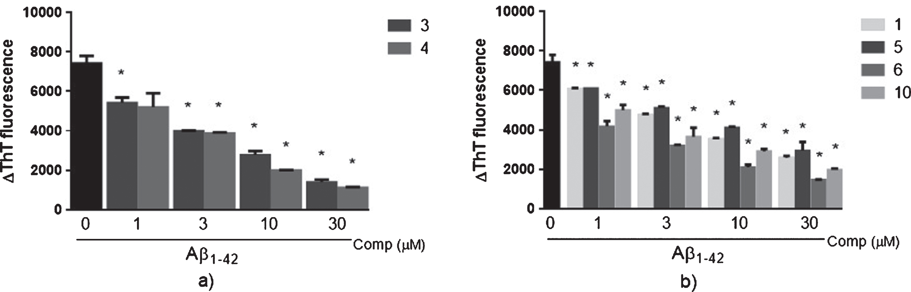
Effect of curcumin derivatives with ether, ester, and diester groups on Aβ aggregation. Aβ42 recombinant peptide was incubated during 48 h with or without different concentrations (1, 3, 10, or 30μM) of compounds. After incubation, ThT (20μM) was added and was determined changes in its fluorescence intensity (ΔThT fluorescence) in the presence of curcumin derivatives with ether and ester groups (a) or with diester groups (b). Results are represented as Mean±S.D. from three independent experiments performed in duplicate. *p < 0.05 relative to Aβ42 alone.
DISCUSSION
New curcumin derivatives were prepared by etherification and esterification in order to provide structural changes that could potentially increase anti-inflammatory activity. Inhibition of the secretion of pro-inflammatory cytokines, such as IL-6, is frequently used as readout of an anti-inflammatory activity. We evaluated the effect of curcumin derivatives on the production of IL-6 by macrophages stimulated with LPS. Assessment of the bioactivity in vitro showed that compounds
Based on the results obtained from the anti-inflammatory activity of curcumin and its analogues, it can be concluded that hydroxyl groups on the aromatic rings of the curcumin are pharmacophores, required for reducing the production of IL-6. Moreover, modifications on curcumin to produce new analogs with potential anti-inflammatory activity by inhibition of IL-6 should take into account the following salient points (1) that at least one hydroxyl group of the aromatic rings should not be modified, and (2) etherification and esterification of only one of the hydroxyl groups present in the benzene rings will strongly enhance the activity depending on the complexity of the substituent added.
At the molecular level, AD is characterized by the presence of extracellular Aβ senile plaques in the brain [1, 5]. Senile plaques are produced by the cleavage of the amyloid-β protein precursor (AβPP) by the enzyme β-secretase at the AβPP beta site, leading to a small soluble AβPPβ fragment and a C-99 fragment of AβPP. The C-99 fragment is then broken by the enzyme γ-secretase into two fragments of AβPP intracellular domain protein and the pathological Aβ42 peptide, which polymerizes forming amyloid fibrils, leading to cell death in the brain [1].
The effect of curcumin on Aβ aggregation has been extensively studied [14, 15]. This effect appears to be favoring the generation of non-toxic Aβ intermediates during the formation of fibrils [16]. It has also been proposed that curcumin disaggregates Aβ fibrils [17]. Compounds
Inhibition of Aβ aggregation is a promising approach for the identification of new agents for AD treatment. Several synthetic and natural compounds have been tested as inhibitors for Aβ aggregation [1]. Some molecules have reached different phases of clinical trials; however, until now, no molecule has been approved as new therapeutic. Hence, the search of new molecules with anti-aggregation effect continues to be of great interest.
Our findings suggest that the novel curcumin derivatives
Footnotes
ACKNOWLEDGMENTS
AAD-A greatly acknowledges financial support from SENACYT (ECS11-002) and INDICASAT-BID (02–12). Financial support to OVL by the Welch Foundation (AX-1788), NIGMS (SC3GM105579), UTSA, and the NSF (CHE-1455061) is gratefully acknowledged. KSR, MG, EM, PLF, and AAD-A would like to acknowledge SENACYT (Panama) and the National System of Investigators (SNI) for supporting their research. JL-B and YG are supported by the Institute for training and Development of Human Resources (IFARHU), and SENACYT (Panama). JL-B is also supported by the Ministry of Economy and Finance (DIPRENA-DPIP-10866-2013) on Nutritive Supplements, and by Melo Brain Grant (Panama). Mass spectroscopic analysis was supported by a grant from the National Institute on Minority Health and Health Disparities (G12MD007591). Authors want to acknowledge Michell Moran and Sahyra Marin for technical collaboration.