
Abstract
BACKGROUND:
Laparoscopic surgeons who regularly perform endoscopy are more likely to develop musculoskeletal disorders than other internal medicine specialists, a difference that attributed to repetitive movements, poor postures, and sub-optimal equipment design.
OBJECTIVE:
This study aimed to design, build, and evaluate an endoscope holder for reducing the static load applied by the weight of the endoscope, in order to reduce musculoskeletal disorders risk factors in the surgeon’s hand, shoulder and back issues regions.
METHODS:
A new endoscope holder was designed according to ergonomic design principles. The designed holder was evaluated by surface electromyography (sEMG) and discomfort assessment including 15 laparoscopic surgeons. The results were analyzed with centrality statistics and compared with the independent t-test using SPSS version 22.
RESULTS:
The evaluation of the new endoscope holder shows a statistical significant decrease in the average electrical activity of biceps brachii, triceps brachii, lateral deltoid, T9 Thoracic erector spinae, L4 Lumbar erector spinae, and external oblique after using the holder (p < 0.05).
CONCLUSION:
The results shows that using the new endoscope holder is associated with a lower level of discomfort, as well as a lower induced muscle activity. The results also highlight the need to upgrade the holder to offer rotability in all directions (perpendicular to the ground) which will be included in the next design.
Introduction
Endoscopy is an essential diagnostic and therapeutic procedure frequently performed in hospitals. While patients’ safety and comfort during endoscopy have been the subject of numerous studies, not much attention has been paid to the safety and health of laparoscopic surgeons who perform this procedure [1–3]. Indeed, it has been reported the patient is not the only person who can be “at risk of sustaining injury during the endoscopy procedure [4]. Numerous studies have shown the high incidence of musculoskeletal disorders in laparoscopic surgeons [5–9]. Ergonomic risk factors associated with endoscopy include repetitive hand movements, excessive hand force, and poor shoulder, neck, and wrist postures [6]. The incidence of overuse syndrome among endoscopists does not appear to be associated with age, gender, or leisure activities. Equipment, and techniques used in this procedure, instead, it is considered an unavoidable consequence [7]. Research has shown that laparoscopic surgeons spend about 43% of their work time performing profession-related activities. It has been reported that laparoscopic surgeons are more likely to have musculoskeletal disorders than other internal medicine specialists, and the risk of musculoskeletal disorders is directly associated with the amount of time they spend performing endoscopy [3, 8]. Given their repetitive movements and potentially harmful postures, endoscopists are at risk of overuse syndrome, resulting in repetitive micro-trauma in connective tissues, and ultimately, collagen deficiency at the microscopic level [10]. Collagen deficiency and trauma in connective tissues can be associated with inflammation, pain, and muscle weakness [10]. This has the potential to give rise to persistent pain and disability [10].
A study by Khanicheh et al. [4] showed laparoscopic surgeons tend to suffer injury and pain in the wrists/fingers, wrists, forearms, shoulders, and back due to poor postures, excessive force, and repetitive movements during endoscopy. While the literature contains several studies on the ergonomic risk factors of endoscopy, few studies have proposed alternative endoscope designs to reduce these risks. In this context, ergonomics conerns understanding how endoscopists interact with endoscopes to perform the intended tasks and how these devices and tasks can be redesigned to minimize the risk of injuries. Endoscopists tend to suffer injuries not because of their weakness or poor training but because of the poor design of the equipment with which they perform endoscopy [9].
The highly reported areas of pain related to Endoscopists are the back, neck, right shoulder, and left thumb [8]. Maneuvers contributing to musculoskeletal disorders among endoscopists include: adjusting tip angulation controls, torquing with the right hand, and standing for prolonged periods [9]. A study by Shergill et al. [7] showed that the age of endoscopists was significantly associated with pain. Although, Shergill et al. reported that the most common painful areas for novice endoscopists were the thumb and fingers (43%), experienced endoscopists were more likely to suffer pain in the left shoulder (33%). Other studies indicated that age and sex were also associated with musculoskeletal disorders [9, 11–13]. Overall, hand torquing maneuvers commonly performed during endoscopy are a significant risk factor for pain. In addition, it has been suggested that left-handed endoscopists tend to feel more pain in the hands and wrists than right-handed ones, although further research is still needed to explore the issue [14]. Mostly frequently of hand movements repeated during endoscopy are radial/ulnar deviation, flexion or extension, pronation and supination respectively [11].
Despite high rates of skeletal disorders reported amongst endoscopists, there is paucity of research into eqiupment design and functionality, more research is needed in the field of design and intervention to reduce these risk factors. Thus, this study aimed to design and evaluate a new holder to minimize this static load to make endoscopy easier and more comfortable to perform and prevent musculoskeletal injuries among endoscopists.
Method
The study includes two main phases: 1) The first phase involved designing an adapted ergonomic holder for the endoscope, using the Schoone-Harmsen method [15]. The method consists of two sub-phases: analysis and synthesis. In the analysis sub-phase, the existing design problems are identified. The synthesis sub-phase concerns providing solutions for the identified problems [16, 17]. 2) The second phase focus on assessing the designed ergonomic holder. The electrical activity of muscles and the level of discomfort before and after using the holder were measured using surface electromyography (sEMG) and the Corlett and Bishop’s body part discomfort scale [15–17], respectively. Electrodes were placed on the belly of six muscles: Biceps brachii, Triceps brachii, lateral deltoid, T9 Thoracic Erector spinae, L4 Lumbar Erector spinae, and external oblique. These muscles were chosen because they play a significant role while working with an endoscope [18]. The bandwidth for recording surface electromyographic waves was 20 kHz/channel (160 Khz total per data LOG). Electrodes were placed on the muscles of the gastroenterologist’s left hand, which upholds the majority of static load from the device during use. Electromyographic signals of each of the six muscles in fifteen subjects were averaged, and the difference between using and not using the holder for each muscle was tested for statistical significance.
Phase I: Ergonomic design and fabrication of an adapted prototype holder for endoscope
According to the studies carried out, musculoskeletal disorders are more prevalent in more experienced (older) endoscopists with heavy daily workloads. Therefore, heavier workloads are associated with endoscopists’ working conditions and need to be countered by an intervention to reduce the ergonomic risk factors of the job. The risk level and increases of musculoskeletal problems increase with body mass index and the number of operations performed per day [7]. Consequently, potential solutions include reducing the number of procedures, prolonging the rest between shifts, performing endoscopy and colonoscopy intermittently, sub-optimal posture throughout the process. Moreover, adding and using an ergonomic holder to improve comfort during endoscopy operations can reduce the strain resulting from the relatively high weight of the endoscope device on the left hand [4, 8]. Before starting the design process, the design team closely observed endoscopy procedures and conversed with laparoscopic surgeons who performed the procedure frequently to determine what would make their job easier and more comfortable. Later on, a prototype was designed to place the weight of the endoscope on the holder, reducing the static load on the left hand, height adjustability at the bottom of the holder, and the connection to the base, in-plane rotability of the panel, and stability of the holder’s base to ensure that it remains still during rotation. After building the prototype, it was decided to modify the proximal component to make it easier to place the endoscope on the holder. The designed holder is displayed in Fig. 1.
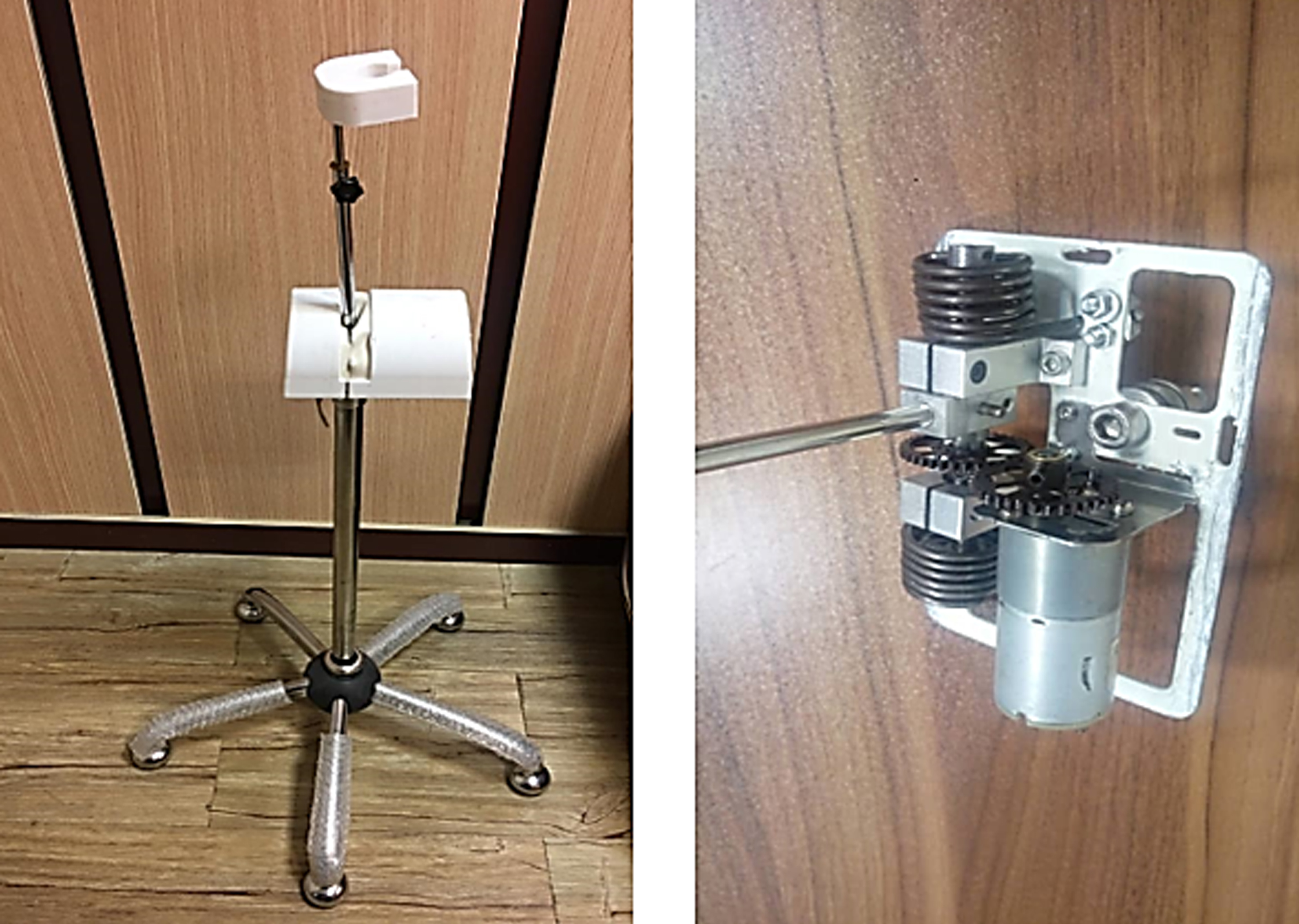
Mechanical components inside the holder (springs act as the lifting force of the endoscope) and prototype of the new endoscope holder.
Participants
Ethics approval was granted by Iran University of Medical Sciences (IR.IUMS.REC.1397.075) and the fabricated holder was tested by 15 (nine men and six women) laparoscopic surgeons (specialists, subspecialists, and gastroenterology fellows) at Firoozgar Hospital. None of the participants had any musculoskeletal pain (in the last twelve months), and all had moderate experience (with almost nine years of work experience) working with conventional endoscopes. All participants filled out the consent form before the test started. The mean age of the 15 subjects was 43.13±10.14 years, and their mean weight and work experience were 72.73±12.72 kg and 8.61±2.72 years, respectively. The subject had at least five years of work experience and did not work as an intern in this medical center. Both left and right handed surgeons were included in our sample.
Surface electromyography (sEMG)
We used the SENIAM electrode (Data Link LS900 apparatus (Biometrics, Ltd, Gwent, UK)) placement standard to record the electrical activity of the muscle superficially (before and after using the designed holder). Since electrodes were wired to the recorder machine, the wires were attached to the body with adhesive bandages to prevent them from moving and causing noise (Fig. 2). The measured electrical waves were recorded on the computer with Data Log software.

Surface EMG electrodes on the participants during work with the new holder. 1: Mechanical holder parts; 2: Location of the endoscope; 3: Holder non-slip bases; 4: Holder height adjustment clamp; 5: Rod connecting the upper part of the holder to the base.
EMG signals needed to be normalized through conversion from absolute value to value relative to a reference point (expressed as a percentage of that reference). The predetermined Maximum Voluntary Contraction (MVC) is commonly used as reference [19]. Therefore, before recording EMG signals, the MVC of each target muscle during the task was also separately recorded. In the process of recording the maximum voluntary contraction, when the muscle bends in the direction of the target joint, the tester resists it, and the electrical activity of the muscle is recorded.
Active electrodes were used for sEMG, and monopolar recording was performed. The sampling frequency is 1200, and the sampling of raw signals is done as a time window.
Discomfort before and after using the designed holder was measured with the Corlett-Bishop scale [15]. This scale measures discomfort in eight regions of the body (neck, shoulders, and arms, back, elbows and forearms, hands and wrists, buttocks, thighs, and knees, and feet and legs) on a scale of 0 to 9, with 0 indicating no pain and 9 indicating maximal pain. This assessment was completed before and after the intervention (twice at the beginning and end of the shift) Assessing discomfort at the beginning and end of the shift was to determine the discomfort acquired elsewhere (not caused by the shift) and remove its effect by subtracting it from the discomfort score reported at the end. For example, for a participant reporting a neck discomfort score of two at the beginning of the shift and seven at the end, the final discomfort score of the shift would be five. This method used in our previous studies and quantified pain and discomfort during shifts (once without the holder and another time with the holder) [16, 20].
Data analysis
The recorded electromyography signals were converted numerically (quantitatively) with Data Log software. The results were analyzed using centrality statistics and a t-test using SPSS software version 22. The paired t-test was used to compare the electromyography and discomfort assessment results obtained with and without the holder. In all statistical tests, p-values of less than 0.05 were considered significant.
Results
The prototype was designed based on ergonomic design principles with emphasis on placing the weight of the endoscope on the holder, reducing the static load on the left hand, and increasing comfort while performing endoscopy without disrupting the routine and with as few changes as possible in the procedure. The ergonomic holder was designed to serve as a holder for the device and as a support for the left hand and transfer the weight of the hand and the device to the base section with sufficient stability to minimize the pressure resulting from the static load of the device on the left hand.
As shown in Table 1, the average electrical activity of biceps brachii, triceps brachii, lateral deltoid, T9 thoracic erector spinae, L4 lumbar erector spinae, and external oblique before using the holder, the t-test showed a statistically significant decrease in the electrical activity of all muscles after using the holder.
Mean±SD scores of electrical muscle activity as a percentage of MVC* during work with the new holder and without the holder (n = 15)
Mean±SD scores of electrical muscle activity as a percentage of MVC* during work with the new holder and without the holder (n = 15)
*Maximum Voluntary Contraction. **Paired t-test.
Table 2 shows the results of discomfort assessments before and after using the holder. As can be seen, after the intervention, the participants experienced significantly decreased discomfort in the neck, shoulders, back, elbows and forearms, and hands and wrists (p < 0.05), but the impact on the hips, thighs, knees, legs, and feet was not statistically significantly (p > 0.05). To estimate the sEMG activity, the values for the total observation time were averaged.
Comparison of body discomfort (10-point Corlett-Bishop scale) in working without a holder and with a holder
*Standard Error. **Paired t-test between before and after using of the holder.
The main goal of this study was to design a new holder for conventional endoscopes to reduce the static load applied to the left hand by the weight of the hand and the endoscope and improve the comfort of laparoscopic surgeons while they perform endoscopy. Our novel ergonomic device was designed for compatibility with all varieties of conventional endoscopes and affordability. By collecting feedback from laparoscopic surgeons during the design phase, the design was optimized to provide comfort during endoscopy and ensure that the wrist posture remains as close to neutral as possible. In addition, the shape of the new holder allows for transferring the weight of the left hand during the procedure, acting as a support for the hand.
After using our designed holder, the participating laparoscopic surgeons reported significantly reduced discomfort in the neck, shoulders, back, elbows and forearms, and wrists and hands. This shows the effectiveness of the intervention in reducing postural pressures on these areas and suggests that it may reduce the prevalence of musculoskeletal disorders in laparoscopic surgeons in the long run. In addition, the ergonomic design principles used in the holder allow it to significantly reduce the pain and discomfort of laparoscopic surgeons in the regions (as mentioned in the result section). However, the holder demonstrated no effec on discomfort in the buttocks, thighs, knees, legs, and feet (which were not the target of the study).
The findings of the present study show discomfort in neck, shoulders, arms, and back among laparoscopic surgeons is hight that are consistent with the results from several previous studies that have reported the highest level of discomfort in the neck, shoulders, arms, and back among laparoscopic surgeons [5, 6].
This study also suggests that the static load applied to the left hand during endoscopy is associated with the potential development of musculoskeletal disorders in laparoscopic surgeons, which is consistent with the findings of previous studies [2, 21]. Accessories that reduce excessive and sustained forces and prevent repetitive movements and non-natural postures during endoscopy can lessen the pain and discomfort caused by performing this procedure regularly [4]. Our findings also indicate that the application of ergonomic design principles in the design of an endoscope holder can significantly reduce muscle activity associated with the reported musculoskeletal discomfort of laparoscopic surgeons and the static on musculature of the hand.
The results of our study confirmed the findings of Boussenna et al., who reported that poor postures of the upper limbs and the duration that the person remains in these postures are associated with considerably increased pain and discomfort [22].
In future research, the performance of the holder can be improved for further improvement in the electrical activity of hand muscles, by making some modifications to the connections so that the laparoscopic surgeons can move the holder with six degrees of freedom (directions x, y, and z plus rotation).
Limitations
Although the present study was a new endoscope holder design, it has limitations. First, the discomfort assessment method was carried out in a short-term trial (one shift of work). Working longer with the new endoscope holder allows participants to familiarize themselves with the new device and provide more complete discomfort feedback. Second, there is an influencing habit with the new device and learning how to work with it. In our opinion, the deployment of such new devices in the endoscopic occupational environment, which is sensitive, should be gradual with more training. Finally, the low sample size, especially in the discomfort assessment phase before and after the intervention, may limit the significance of the study results. In similar studies, the low sample size is often due to the difficulty of obtaining the consent of the participants to participate in the study due to lack of familiarity with new system [23, 24]. Participants in our study are experts in their professions and volunteer to participate in the study, and a compromise that often must be made is to limit the number of study participants. Future work should include more validation studies in a larger sample size.
Conclusion
We discussed in this paper our attempt to design and evaluate an endoscope holder for reducing the static load applied by the weight of the endoscope. The evaluation phase showed that using the new designed holder reduces the static load and pressure applied to the lateral deltoid and biceps brachii muscles of participant laparoscopic surgeons during endoscopy. The reduction of the static load also decreased the level of user-reported discomfort in the hand after multiple endoscopy procedures. In addition to reducing the static load, the designed holder also transfers the weight of the hand to the ground, which results in reduced pain and discomfort in the upper body that contributes to withstanding static loads during endoscopy (wrist and hands, elbows and forearms, back, shoulders, and neck).
Ethical approval
This study was approved by the National Committee for Ethics in Medical Research (IR.IUMS.REC.1397.075). All procedures for studies involving human participants were performed in accordance with the ethical standards of the institutional and/or national research committee.
Informed consent
All participants filled out the consent form before the evaluations.
Conflict of interest
The authors have no potential conflict of interest to disclose.
Footnotes
Acknowledgments
The authors would like to express their sincere gratitude to the expert pannel in this study.
Funding
This research was supported by Iran University of Medical Sciences (IR.IUMS.REC.1397.075).