
Abstract
In this paper, we present a review of the research literature regarding applying X-ray imaging of baggage scrutiny at airport. It discusses multiple X-ray imaging inspection systems used in airports for detecting dangerous objects inside the baggage. Moreover, it also explains the dual energy X-ray image fusion and image enhancement factors. Different types of noises in digital images and noise models are explained in length. Diagrammatical representations for different noise models are presented and illustrated to clearly show the effect of Poisson and Impulse noise on intensity values. Overall, this review discusses in detail of Poisson and Impulse noise, as well as its causes and effect on the X-ray images, which create un-certainty for the X-ray inspection imaging system while discriminating objects and for the screeners as well. The review then focuses on image processing techniques used by different research studies for X-ray image enhancement, de-noising, and their limitations. Furthermore, the most related approaches for noise reduction and its drawbacks are presented. The methods that may be useful to overcome the drawbacks are also discussed in subsequent sections of this paper. In summary, this review paper highlights the key theories and technical methods used for X-ray image enhancement and de-noising effect on X-ray images generated by the airport baggage inspection system.
Keywords
X-ray imaging
X-rays are in fact a type of waves like electromagnetic radiation which is commonly used for taking images of a body structure. It is just like other visible light, radio waves and gamma rays but comparatively more energetic and highly penetrating form of radiations. It can pass through several materials and the image is recorded on computer using the under mentioned ways [1]; Dense structures such as bones or metals block most of X-ray photons and appear as white. Structures such as air appear as black and Structures like fat, liquid and muscle appear as shades of gray.
Photons can pass through the low density materials directly, however higher density materials reflect or absorb the photons because there is a smaller amount of space between the atoms for the waves to pass through [1]. X-ray imaging is a well-established technology and is widely used in different areas like aviation security and medical imaging. Dr. Roentgen, professor of physics, is founder of discovering X-ray in 1895 [2]. X-ray imaging works by placing an object between the X-ray source and the receptor (a film or a detector). The exponential law for a narrow spectrum beam explains the physics of X-ray imaging is presented in equation 1 [1, 3].
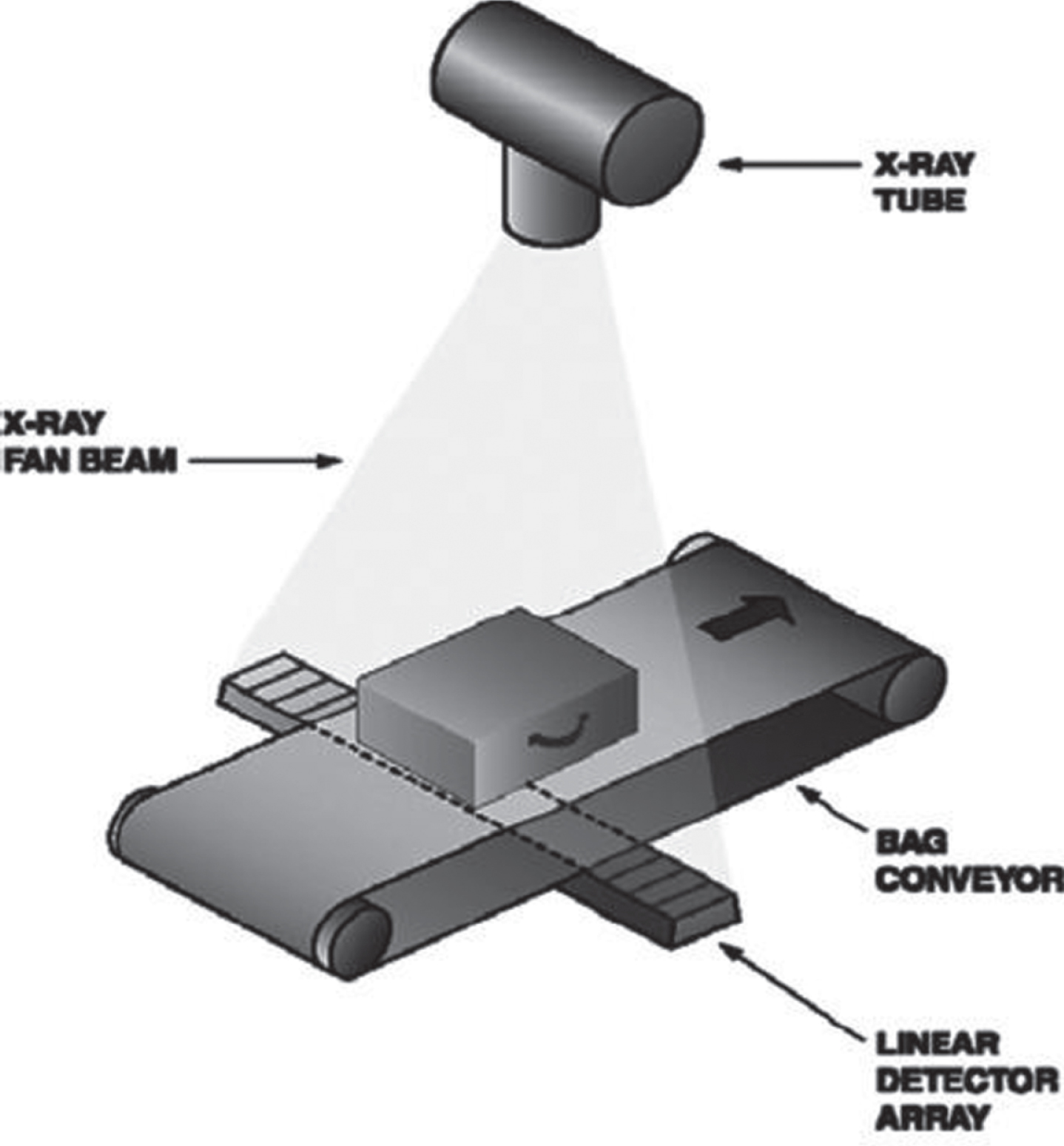
A typical X-ray imaging system has a source that generates X-ray photons; a detector array, usually L-shaped which records the X-ray photons; and a signal processor unit performing signal and image processing computations such as analog to digital conversion, contrast enhancement, noise reduction or other higher level image processing. The operator display presents the output to the users. Figure 1 shows a simple dual energy X-ray inspection system. The physical principles of X-ray baggage inspection can be used to interpret the results and to determine the type of materials exist in the object [4]. X-ray screening approaches are commonly used at airport to inspect baggage for security purposes. X-ray technology provides detail information about the density of the object denoted by d and the effective atomic number Zeff. During X-ray scan, an object’s material type can be determined using its density and atomic number [5]. X-ray technology is safer than nuclear magnetic resonance both to human beings and to the contents inside the baggage. X-ray physics is easier to operate and cheaper than the neutron-based approaches.

Dual energy X-ray inspection system [6].
Baggage inspection system plays an important role at airports. Different techniques have been employed to detect illicit objects or contrabands and explosives [7]. These techniques are detailed in the following subsections.
Conventional transmission x-ray system
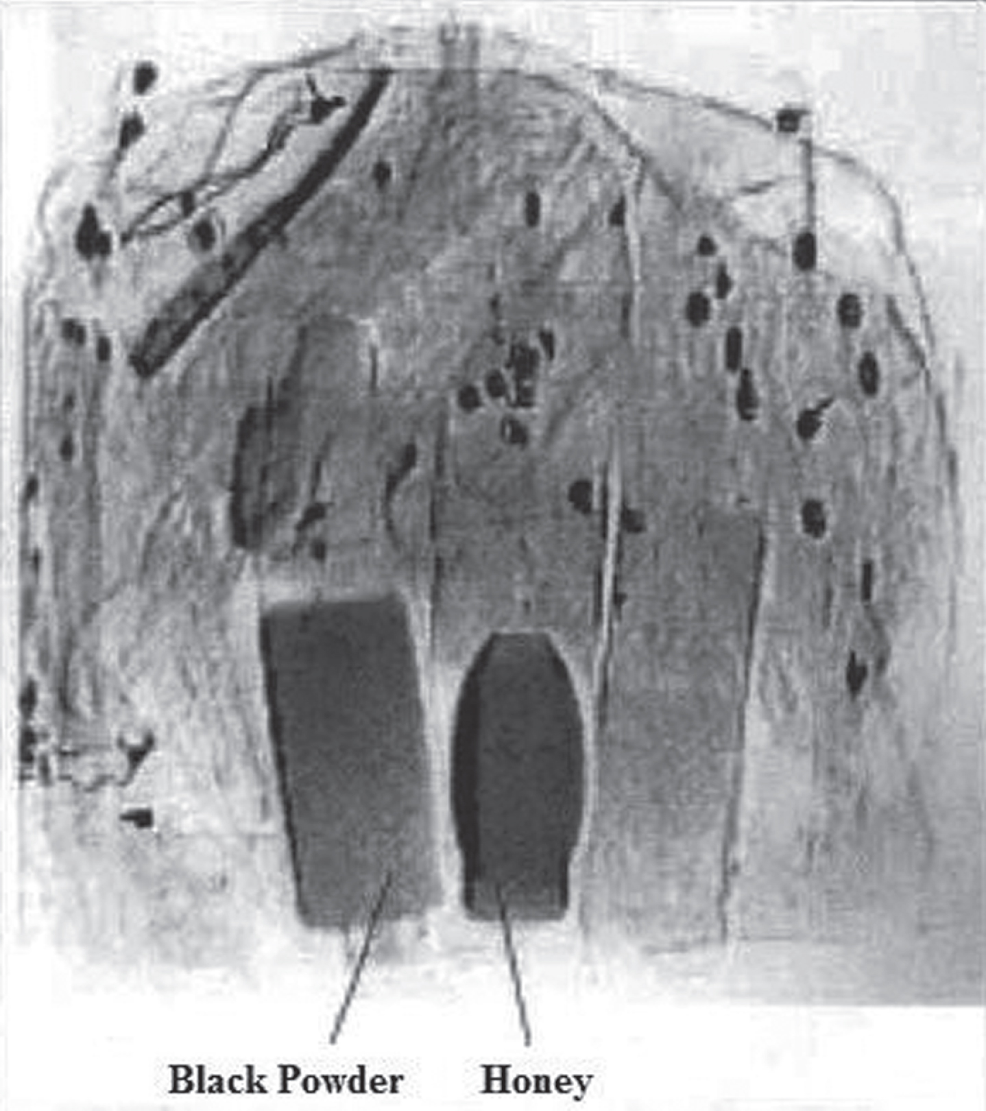
X-ray technology is widely used in the aviation manufacturing for illicit material or threats detection [38]. Conventional transmission X-ray imaging system or single energy X-ray system has a scanning X-ray beam that is transmitted through the baggage meant for scrutiny. A line of detectors measures the absorption of the X-ray and a high-resolution X-ray image, derived from the degree of absorption of the X-ray generated. Figure 2 depicts a commercial conventional transmission X-ray carry-on baggage detection system. The advantage of the single or conventional energy inspection system is its high speed. It is capable to check up to 1500 baggage in an hour. However, the conventional energy scan has certain limitations. Like X-ray shadow produced by black powder, it will resemble to the shadow of honey as shown in Fig. 3 [8]. Consequently, it is possible to discriminate among dissimilar density materials using X-rays with two different average energies [9]. To eradicate the problem, dual energy X-ray systems were developed. It improves the characteristics of single energy systems and that is why dual energy X-ray systems are widely used at airports [10, 11].
Conventional X-ray inspection system [71].
Discrimination by shape is a big problem [11].
In the detection of illicit objects, a dual energy X-ray inspection system uses two types of X-ray energy levels, i.e. low energy and high energy. In this system, high energy over 100 kV is used to detect object density. It absorbs energy which primarily depends on the density of the material. The higher is the density, the more will be the energy absorbed by the object and consequently the darker the X-ray image [12]. At lower energy levels, around 50 to 80 kV, the absorption depends on the effective atomic Zeff number as well as the thickness of the material. That’s why the areas of heavy metal will be dark in both low energy and high energy X-ray images and areas of light elements are darker in the low energy X-ray images only [13]. Light elements such as carbon, nitrogen, and oxygen can be detected by comparing both images [14].
The problem of differentiating between a thin sheet of strong absorber and a thick slab of a weak absorber, faced in single energy X-ray imaging systems, has been solved in dual energy X-ray baggage inspection systems. However, the accuracy of estimating the effective atomic number of a material in baggage is still uncertain because possible false alarm rates are as high as 20% or more [13–16]. The X-ray images produced by the inspection system are still noisy, blur and with low contrast. Dual energy X-ray system enhances the functionality of single energy X-ray system. Therefore, this system is widely used at airports [10, 16].
X-ray CT (computed tomography) scanner system
The main characteristic of X-ray CT scanner is that it develops detection accuracy. Figure 4 shows a typical airport CT scanner. It produces virtual slice or tomography images of specific area of the inspected object using processed X-ray. The CT inspection system uses cone beam X-rays in which the baggage remains stationary and a huge area of X-ray detector, which covers the entire baggage, takes the images. It can collect about 360 images in one rotation and can get 3D images of the objects using reconstruction software. In this system, the contrast between objects inside the baggage does not depend on the thickness of the object but it primarily depends on the type of material, that’s why, it is a worthy tool for discriminating between different materials [10]. However, taking CT images and reconstructing them is a time-consuming task as compared to single and dual energy X-ray images. It is not convenient to check every piece of baggage [10]. CT inspection system is only used as a second layer and only checks the selected baggage identified by first layer with X-ray inspection systems.
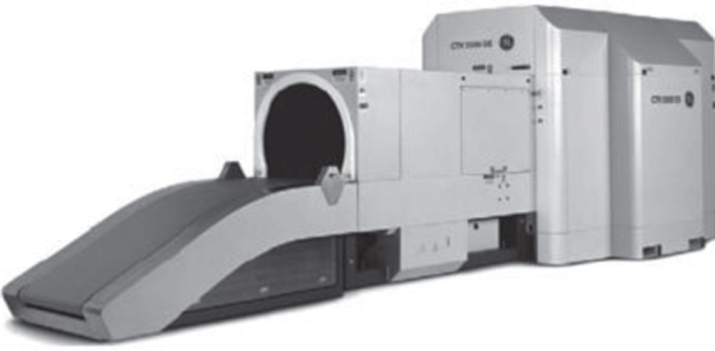
Computed tomography scanner [10].
The CT scanner can completely separate the superimposed objects and can differentiate between materials with different photons absorption. However, since it detects threats in similar absorption coefficient; it will be quite difficult to distinguish between a fruit cake and explosive plastic in sheet form [10].
X-ray backscatter is an advance technology. It detects the radiation reflects from the target and forms an image. It is efficient for inspecting organic materials. These systems were basically developed for inspecting the people from head to toe for searching illicit or contraband items in a person’s clothing, inside the human body and his artificial limbs [17, 18]. Figure 5 shows a backscatter low dose body inspection system. This system can be used for large objects like trucks and containers. Normally it is used for corpse checking and not for baggage inspection.
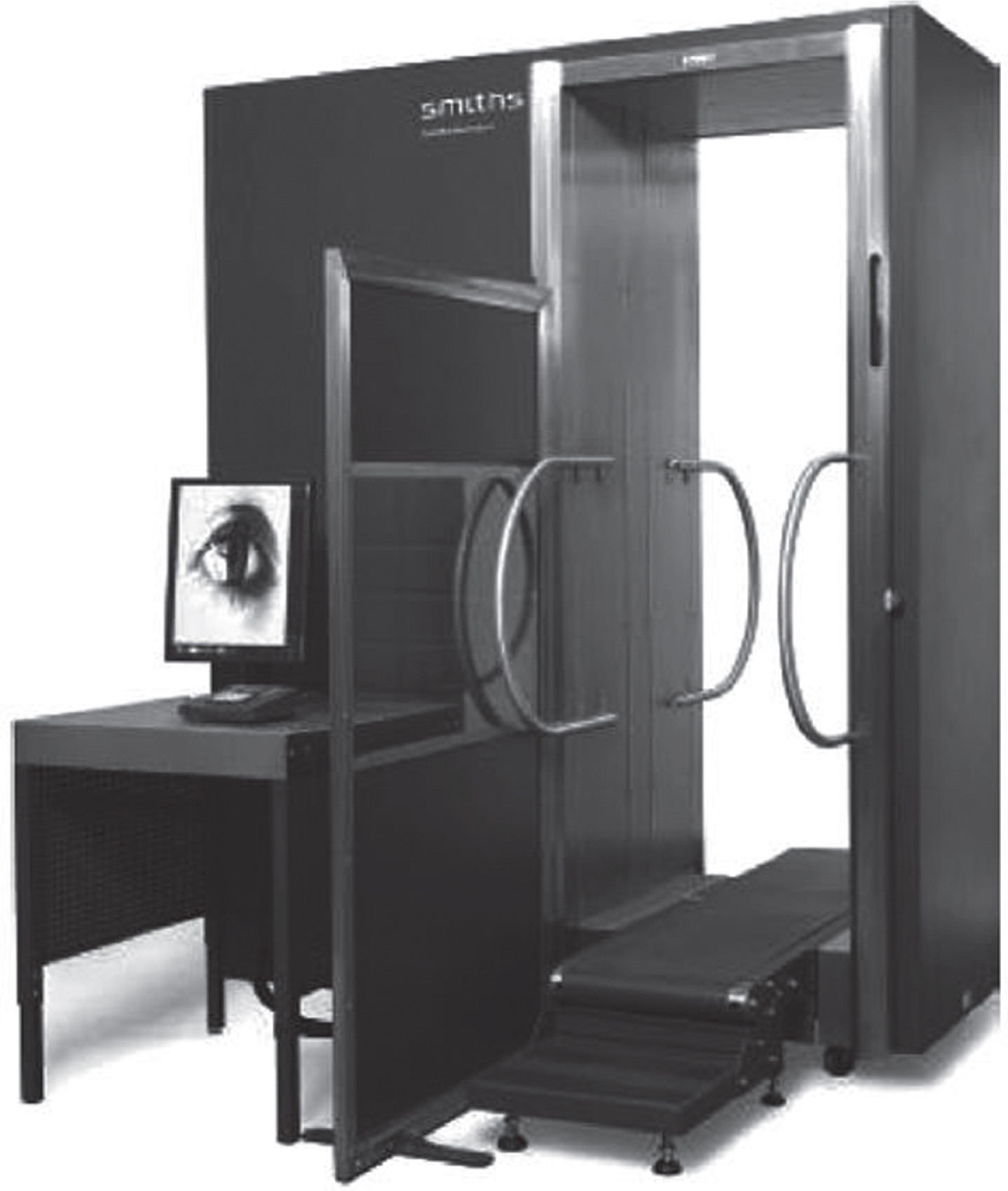
Photograph of the low dose body scanner [18].
There are many other X-ray inspection systems suitable for security screening, but these are very time consuming, difficult to operate and expensive. Some of these are Nuclear based explosives detection systems like Fast Neutron Analysis (FNA), Thermal Neutron Analysis (TNA), Pulsed Fast Neutron Analysis (PFNA), Pulsed Fast Thermal Neutron Analysis (PFTNA), and Nuclear Resonance Absorption (NRA) of gamma rays [12].
Section 2.2 clearly indicates that dual energy X-ray imaging system uses two X-ray energy levels, i.e. low energy and high energy, for the detection of weapons and contrabands. The most important thing is the fusion of these two dual energy X-ray images. The aim of image fusion is to integrate the meaningful and similar information from both the high energy X-ray image and the low energy X-ray image so that the resulting fused X-ray image is more amenable for a successful screeners’ interpretation [13]. Figure 6 shows X-ray image fusion process.
X-ray image fusion process.
Normally dual energy X-ray images can be fused by using two types of techniques [19]. Image Fusion using Local Spatial Information Image Fusion using Wavelet Transform
This fusion technique highlights such details which are out of sight or not clearly seen in original dual energy images using the following steps. Find the difference image ‘Diff’, using equation 2.
For image differencing, classify every pixel in the baggage scene as a detail pixel or local background pixel. Here a threshold value is used to differentiate background pixels and detail pixels. The background image and detail image can be calculated in a simple way using equations 3 and 4.

Dual energy X-ray image fusion using local spatial information [19].
The technique of fusing X-ray images based on ‘wavelet transform’ is presented in Fig. 8. First, Discrete Wavelet Transform (DWT) is applied to low energy and high energy X-ray images to achieve approximate coefficients and detail coefficients. Primarily, a wavelet family and a wavelet basis proficiency of representing X-ray image information need to be selected. In X-ray image fusion process, the wavelet family and wavelet scales will affect the decomposition of coefficients. Because fewer scales will cause the loss of details in the fused X-ray image but too many scales will result in a rough fused X-ray image and may cause difficulties for screeners to understand [13]. The research work used “Haar” as a wavelet family” due to its simplicity and easy to use and “4” level of decomposition is used as a scale. There is no need for multiplications. It requires only additions and there are many elements with zero value in the “Haar” matrix, so the computation time is short. Similarly input and output length is the same that’s why widely used in literature.
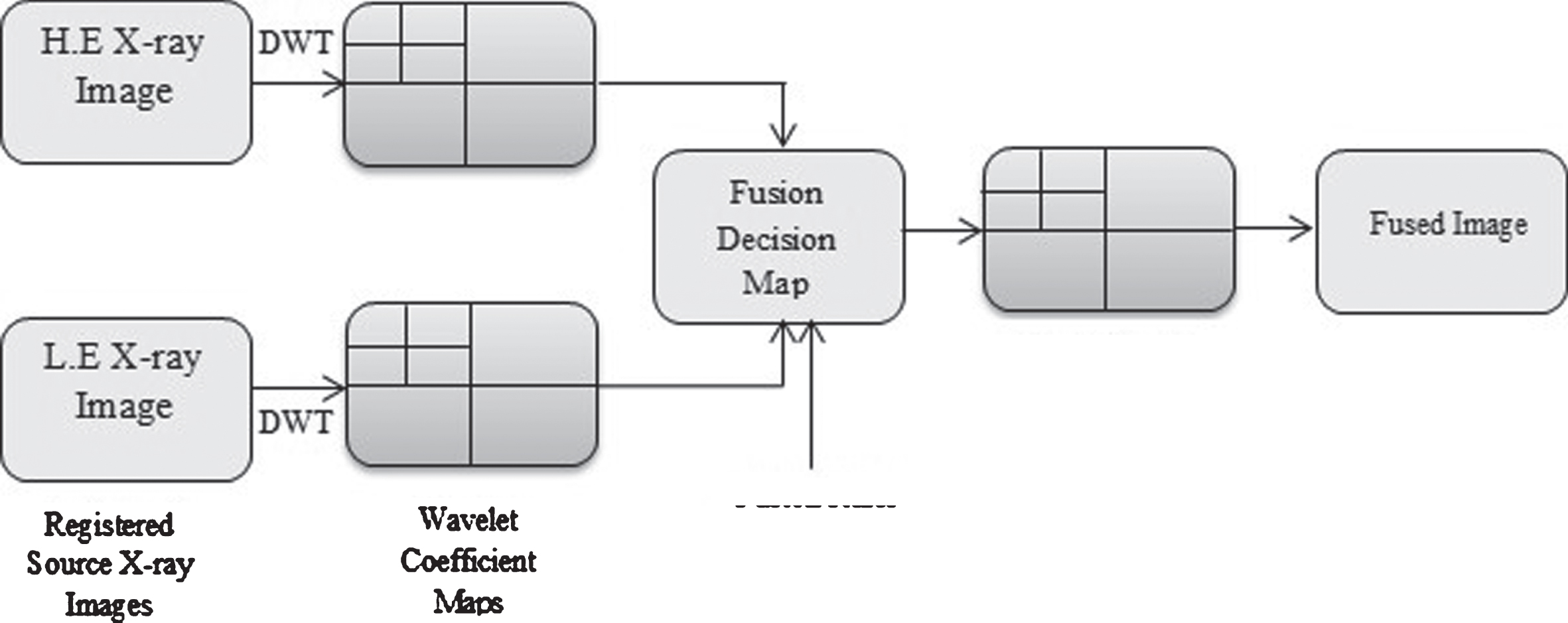
Dual energy X-ray image fusion using discrete wavelet transform [20].
In wavelet-based X-ray image fusion, each of the input X-ray images are transformed into conforming wavelet coefficient X-ray images using the DWT. DWT is normally used for image fusion [15, 21]. The fused X-ray images are decomposed to calculate its approximation and details coefficients. These coefficients can be calculated using low pass filter. Then, using a fusion instruction, the fused wavelet coefficients are computed from the wavelet coefficients of the source X-ray images. At the end, the inverse discrete wavelet transform (IDWT) is applied to the fused wavelet coefficients to obtain the fused X-ray image [13].
This study used dual energy Baggage X-ray images. Therefore, we will fuse those images to make it suitable for post processing steps. Image fusion using DWT is a simple and widely used approach in the literature.
The interpreter or screener cannot read a raw image directly obtained from inspection system. There are some factors, which must be handled properly to get proficient results. These quality factors are: Image Noise Image Blurriness Image Contrast Image Artifacts Image Distortion
Reasons stated above are the key factors for image quality enhancement. However, this study is an effort to improve feature enhancement especially Poisson and Impulse noise reduction due to which there are still 20% or above false alarm rate [10, 13–15]. In the next subsection, brief explanations for these image quality factors are presented. The study introduces the above stated image quality factors, but it has the detail explanation about noise factor in Section 5. Figure 9 illustrates image quality factors.

Image enhancement quality factors.
Image noise is the most important factor that degrades image quality. Image noise is the unwanted information in images. It is a random variation of color information or brightness in images. Different factors like camera illumination sensor, circuitry of scanner or transmission and conversion from signal to digital can cause image noise. Noise effected images can be mottled, grainy, textured, or snowy in appearance. The different types of noises are Gaussian noise, Impulse noise, Poisson noise, Speckle noise, Film grain etc.
Blur effect on images
Image blurriness is another important factor that degrades image quality. The key effect of image blur is to decrease the contrast and the visibility of small objects in images [22]. Figure 10 depicts the blur effect on image. It is apparent that the boundary between visible and invisible objects is determined by the contrast sensitivity of the inspecting system. It has a minor effect on the visibility of large objects, but it decreases the contrast and visibility of small objects [22]. Image blurriness occurs due to various reasons. One of these is the cause of image noise removal, which this thesis also aims to resolve. The objective achieved in this thesis is to reduce the noise in such a way to prevent image blurriness. Dual energy X-ray images also have a severe problem of image blurriness because the object is inspected on a moving belt of the machine.
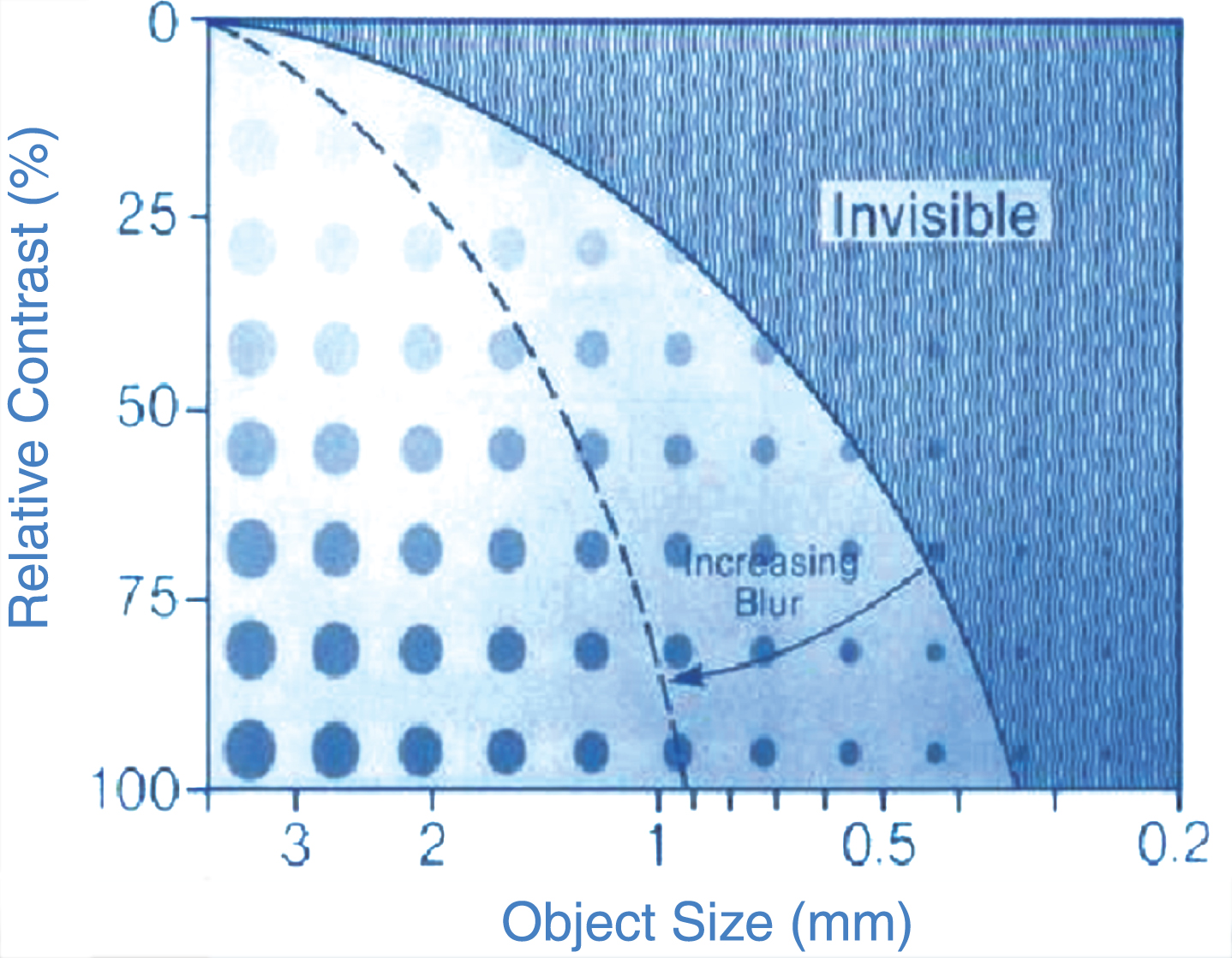
Blurriness effect on images [22].
Another factor that degrades image quality is image contrast. Image contrast can be in the form of various colors, shades of gray or light intensities. Basically, contrast is the amount of difference between tones in an image. An image may have both high contrast and low contrast. Various approaches are used for image contrast like histogram equalization, contrast specification, un-sharping etc.
Artifacts effects on images
Image artifacts are another issue which makes certain image structures that do not appear in an object of interest. In many situations, image artifacts do not considerably affect object visibility and accuracy. However, it can vague a part of an image [22].
Distortion effects on images
X-ray image should not only make internal objects visible, but it gives a correct impression of their shape, size, and relative positions. Distortion is the degradation in image shape, size and positions. An imaging technique can, however, introduce distortion of these three factors [22].
Digital image noise types
Image noise is the most important factor among the five image quality factors discussed early that degrades image quality. Image noise is the unwanted information in images. It arises due to the sensor and circuitry of a scanner or digital camera or due to conversion from signal to digital format [23]. Below are the detail explanations about digital image noise types.
Gaussian noise
The typical model of amplifier noise is additive, Gaussian, and is independent at each pixel and the signal intensity as well [24]. Gaussian noise usually arises during acquisition process like high temperature or poor illumination or transmission. You can see and hear Gaussian noise when you tune a television channel. The pink and the white noise are the Gaussian noise. In color cameras, there is extra noise in the blue channel as compared to green channel [25, 26].
Impulse noise (salt & pepper noise)
Another type of noise, which occurs in digital images, is Impulse noise. An image having Impulse noise will have bright pixels in dark regions and dark pixels in bright regions [23]. Impulse noise usually arises due to dead pixels, bit errors in transmission and analog-to-digital converter errors. It can be eliminated in large by using interpolating around bright or dark pixels and dark frame subtraction. Impulse noise models and its filtering methods are discussed in detail later in this study.
Poisson noise (random noise or photons noise)
The most important noise is Poisson noise. It usually occurs in X-ray images [27, 28]. The reason for Poisson noise creation is due to less penetration, randomly dropping of photons, and size of detector. Poisson noise also arises when the finite numbers of photons which carry energy are small enough to give rise to detectable statistical fluctuations in a measurement [29]. These particles are called electrons in an electronic circuit or photons in an optical device. Detail discussion about Poisson noise is in Section 6.
Speckle noise
Speckle noise normally arises in ultrasound and synthetic aperture radar (SAR) images and it degrades the quality of those images. Speckle noise in SAR is very thoughtful and creates problems in image interpretation. It is like light and dark pixels in radar waves. Speckle noise arises from the signals of elementary scatters, the gravity-capillary ripples, and manifests as a pedestal image under the image of the sea waves. One method, for example, employs multiple-look processing [30]. It can be simply reduced by spatial filtering and multi-look techniques [31]. Normally, speckle noise can be reduced after image acquisition process.
Film grain
The Film grain is a spatially correlated and temporarily independent noise. This noise is very much prominent in higher resolution images. Similarly, it is noticeable in high definition videos. In the areas where the probability is low then the distribution will be close to the classic Poisson distribution of shot noise. However, this noise is very difficult to encode due to its random characteristics. Furthermore, it also affects the accuracy of motion estimation.
Impulse and poisson noise models
In this section, first we will discuss the different Impulse and Poisson noise and then represent diagrammatically all these noise models and its effect on intensity values of X-ray images.
Noise model 1
In noise model 1, X-ray image grayscale values are corrupted by two static values “0” or “255” generated with the same probability. i.e. if pixel value in an image at location (i, j) with grayscale value s i ,j, then the corresponding pixel of the noisy X-ray image will be x i ,j, in which the Probability Density Function of xi,j is:
In this technique, the density of noise within the window can be calculated by equation 7 where N A represents the number of impulses and N represents the total number of pixels. It can express mathematically as below:
Noise model 2
Noise model 2 is very much related to noise model 1, however, each grayscale value might be degraded by either “0” or “1” salt or pepper noise with unequal probabilities [33, 34]). Equation 8 illustrates noise model 2.
Noise model 3
In this model, instead of two fixed values, the grayscale values are corrupted by two fixed ranges that appear at both ends with a length of m each, respectively. For example, if m is 10, then the grayscale value can get any value in a range of (0, 9) or (246, 255) [33]. Equation 9 illustrates noise model 3.
Noise model 4
Noise model 4 is related to Model 3 and the grayscale value is corrupted by two fixed ranges but with unequal probabilities as described in noise model 2. That is:
Noise model 5 (Poisson noise)
Poisson noise can affect X-ray image pixels intensity value with any random value of m with respect to the neighboring pixel i.e. pixel can get any random value. Equation 11 illustrates noise model 5.
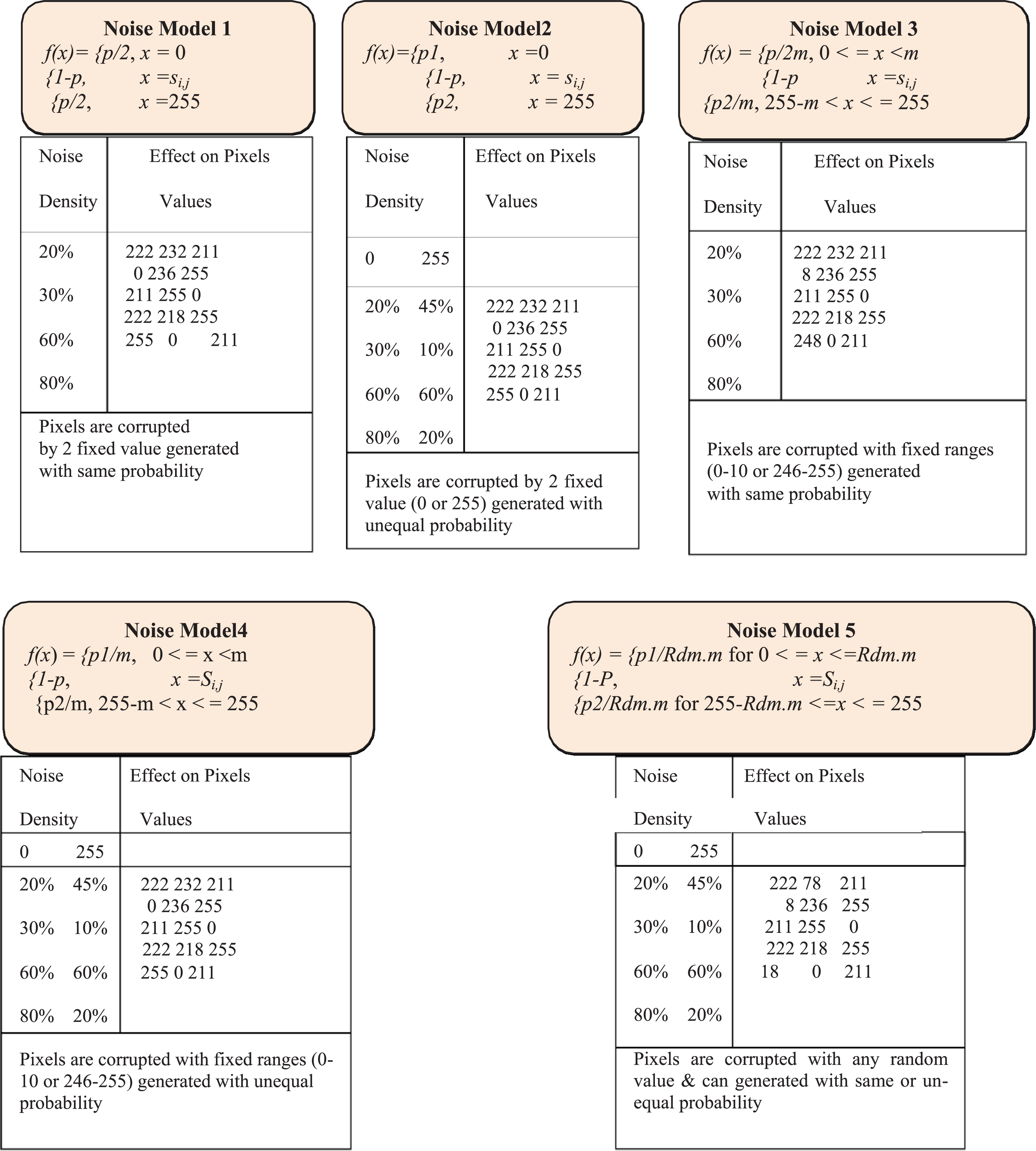
Noise models effect on pixels intensity values.
In Fig. 11, five noise models are presented with its effect on image intensity values. Noise models 1 and 2 relate to Impulse noise as explained in section 6. It affects image pixels values either by “0” or by “255”. Similarly, noise models 3 and 4 also relate to impulse noise effect on image pixels values with a random value in a fixed range (i.e. 1-10 or 246-255). It means that the pixel value will be corrupted by a random value ranging from 1-10 or 246-255. The last noise model 5 relates to Poisson noise, which normally exists in X-ray images. It affects image pixels values by any random value, which means that it affects the intensity values of a pixel due to sudden intensities of the neighbor pixels. In this mode, the X-ray image intensity will be corrupted by and random value that can be 8, 15, 0, 255, 84. The above stated models appeared in single energy X-ray images and dual energy X-ray images.
X-ray images normally contain Poisson noise or source noise. In this section, the basic reasons of Poisson noise and its effect on X-ray images will be discussed. Poisson noise arises when the finite numbers of photons carrying energy are small enough to give rise to detectable statistical fluctuations in a measurement [29]. These particles are called electrons in an electronic circuit and photons in an optical device. Unlike others digital images, X-ray images usually are degraded by Poisson noise and some time by Impulse noise. Most of the previous research work is normally on Impulse noise removal and research studies introduce very efficient techniques for Impulse noise elimination but are not trying to mitigate Poisson noise problems in spatial domain (Pixel by Pixel operations). Some research studies have worked on Poisson noise in frequency domain [27]. They proposed a framework to reduce Poisson noise using Wavelet Transform and Modified Bayes-Shrink method in wavelet domain. The research work in [35] also tried to remove Poisson noise from X-ray images using Wavelet domain. Detail explanation about the reasons of Poisson noise on objects in X-ray images and its effect are discussed in the next sections.
Reason for poisson noise
X-ray images are contaminated by Poisson noise, photons noise or shot noise and sometime called as random noise. Randomly dropping of Photons, less penetration of photons, thickness of the object in image, size of detector problem and when the finite numbers of photons carry energy are small enough to give rise to detectable statistical fluctuations in a measurement are the basic reasons which create Poisson noise in X-ray images.
Below section “a” and “b” presents detail of randomly dropping and less penetration of photons. X-ray photons from the X-ray beams drop randomly on inspected object. Therefore, some areas of the object under inspection get more photons while the other gets less. This discontinuity creates Poisson noise in X-ray image. Figure 12 expresses the matter diagrammatically.
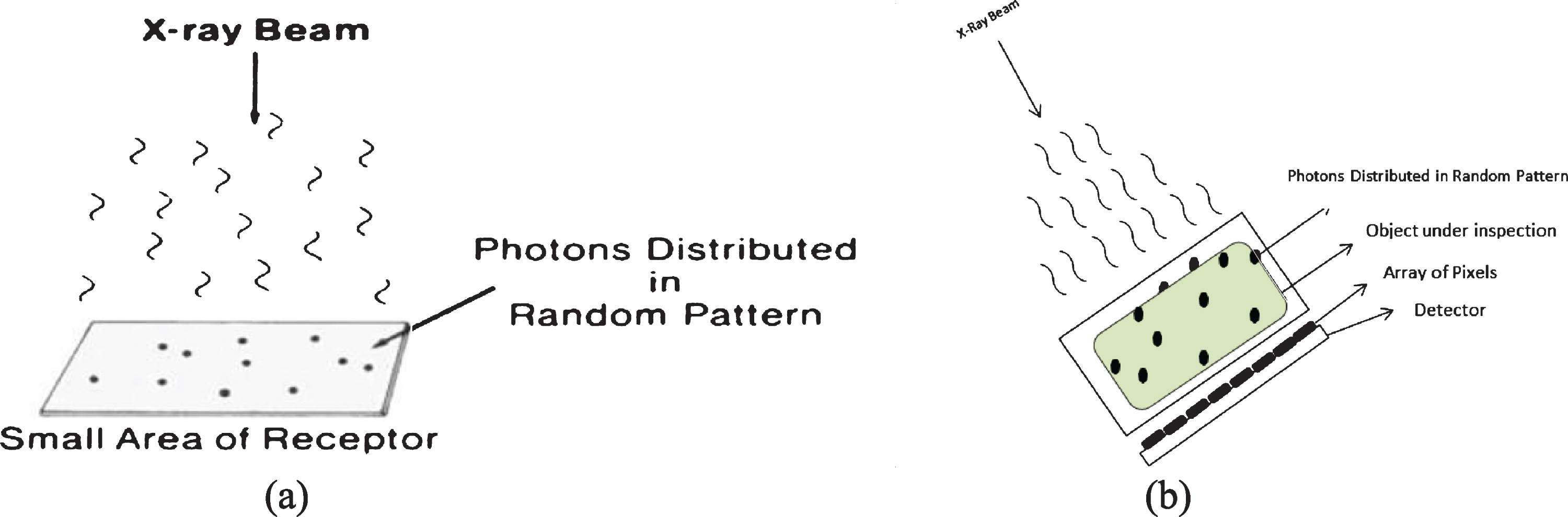
Randomly dropping of photons.
Detail explanation of Figs. 13 and 14 has presented thickness & detector size problem. The X-ray sources usually produce a fan-shape beam which means that pixels collect X-rays from various viewpoints or angles. Figure 13 depicts the variance which arises in X-rays coming from various ways. When the X-rays are coming from same direction to the surface of a thick object with thickness t, the detectors will read and measure the similar thickness from all three X-rays. However, when the X-rays are coming from different directions, the detector will measure different readings [36]. If an object collects X-rays from different directions, there will be an obscurity effect around the edges of an object. The detectors size will also affect the edges because actually the pixels are shaped by averaging the pulse sample within the length of a detector. This effect is much prominent when detectors are larger [36].
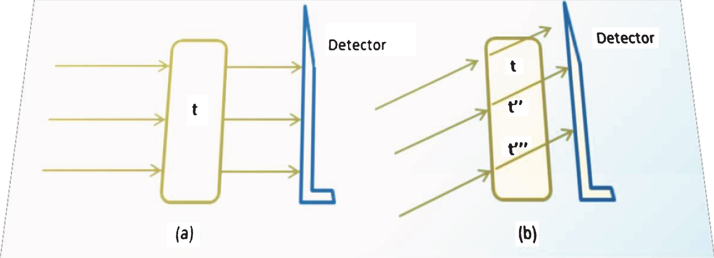
Object thickness problems [36] – Edge effect shows (a) when three similar X-rays anticipated on an object having thickness t from same direction, detectors will have the similar measures for all X-rays. However, in diagram (b) when the X-rays come from different directions, detectors will have different measures due to the thickness of the object.
As illustrated in Fig. 15, an object with thickness t and length equivalent to three detector width will not have much misreading when the signal from the object is evenly distributed to all three detectors as shown in (a). It will be read as 4 pixels wide when the object is not properly aligned with the detectors as shown in (b). Detector 3 and detector 4 will still have the reading t, while detector 2 and 5 will impress from the object and the air. The pixel value of detector 2 and detector 5 will neither be t nor zero, rather it will give uncertain value. The output image will be a 4-pixel-wide object with 2 pixels in the center darker than the edges. The problem can be minimized by increasing the resolution. When the detector size becomes half of its original size as shown in (c), 6 pixels will have the same reading while detector 1, 2, 3 and 4 will have different readings. Thus, the above basic reasons created Poisson noise in X-ray images that lead to un-certainty for the system while discriminating objects and for the screener in decision making process. In the next section, the research work will discuss the effect of Poisson noise on X-ray images.
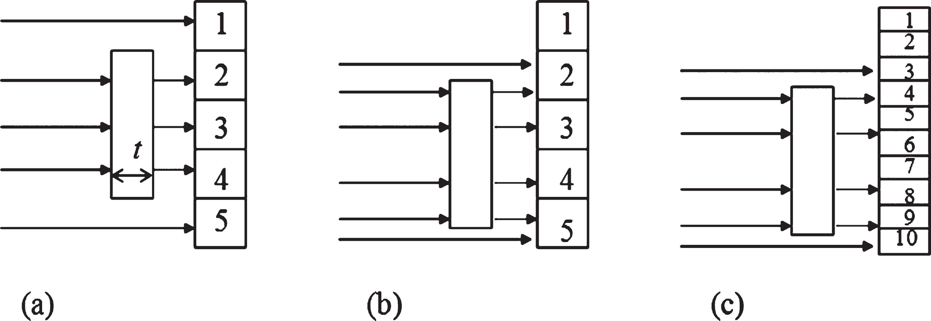
Detector size problems – Edge artifact and resolution illustrate (a) an object with thickness t and length equivalent to three detector’s size is placed aligned with detector 2, 3, and 4. They will have the same readings. (b) The edge of the object falls within detector 2 and 5; detector 3 and 4 will have different readings than detector 2 and 5. (c) When detector size becomes half of the original one, most parts of the object fall within detector 5, 6, 7, 8, and 9.
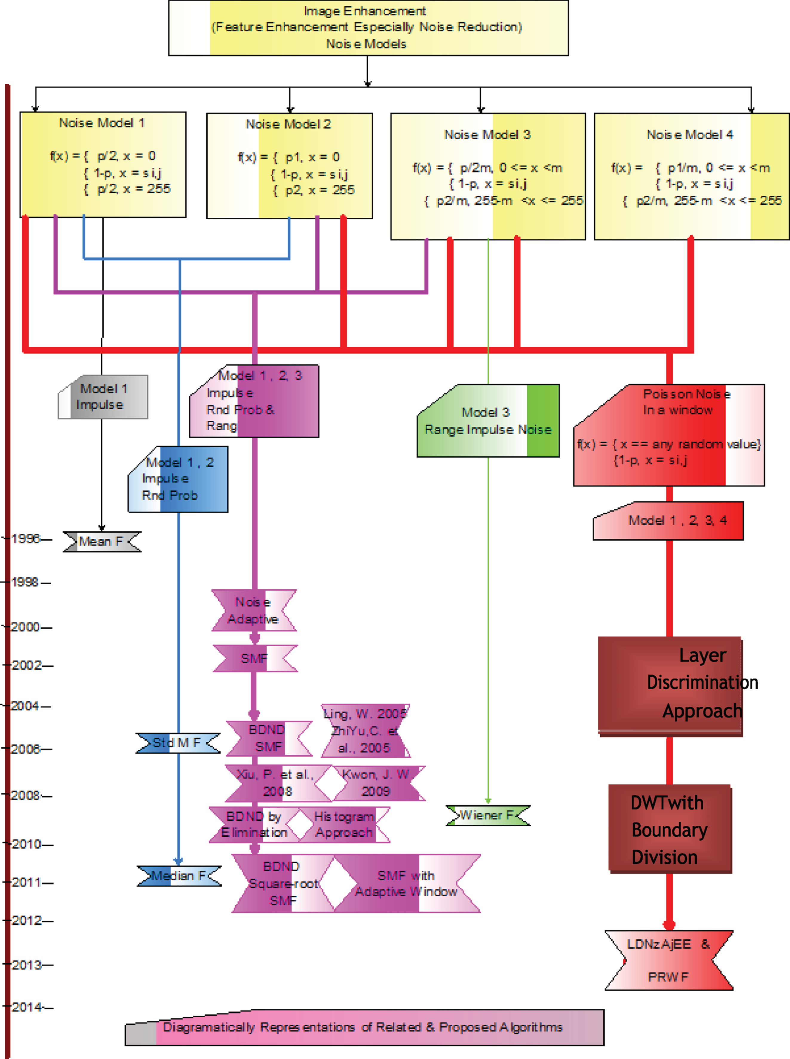
Diagrammatically represents related and proposed work with noise models.
The effects of Poisson noise on X-ray images are: Poisson noise damages the edges of objects in X-ray image which create uncertainty for the system while discriminating objects in X-ray image, Poisson noise affect the pixels value which intensities vary abruptly from the intensities of the neighboring pixels, and makes screener’s decision difficult whether the alarm is false or true.
Above stated effects are the basic problems in X-ray images caused by Poisson noise. This thesis design and formulate new approaches to mitigate these problems. The next section discusses different approaches used for X- ray image enhancement.
Image quality measurement factors
Image quality can be measure using image quality statistical measurements factors. Peak-signal-to-noise-ratio (PSNR) and structural similarity index measure (SSIM) are the most frequently used criteria in the literature to measure the quality and performance of any de-noising algorithm quantitatively [37, 38]. Thus, the performance of the SSIM is computed as:
Similarly, we can use SSIM to assess the similar details between original image and restored images. SSSIM can be calculated using equation 14.
Higher PSNR value represents the good the image quality and the image quality decreases with lower PSNR values. Similarly, SSIM with lower value shows that the original and restored image has similar details information and SSIM with lower value represents the loss of details.
In the literature, processing of digital images and X-ray baggage images have been made progressive through several methods. Different approaches often use traditional methods like image enhancement, de-clustering, de- nosing and pseudo-coloring algorithms. The idea presented in [39] proposed an image enhancement approach using Logarithmic Transform Histogram Shifting (LTHS) algorithm but this approach was only useful in Impulse noise reduction. The theory of [40] used techniques like Contrast Stretching, Logarithmic Intensity Adjustment, Gamma Intensity Correction, and Histogram Equalization for image enhancement. Similarly, the approach in [19] described a dual energy X-ray image fusion approach using Local Spatial Information approach. This approach first classified each pixel in a baggage scene as a background pixel or detail pixel, and then highlights detail pixels with the postulation where they transmit the features of interest in the baggage scene. The idea in [37] used a conventional Mean filter for Gaussian noise removal and did not present any approach for Poisson noise removal. The idea in [24] used Median filter, Mean filter, Wiener filter for Impulse noise, Gaussian noise, Speckle noise, and Poisson noise reduction. The idea presented in [41] used Median filter for Impulse noise reduction. Similar work presented in [42] used Weighted Median filter and the work in [43] used Center Weighted Median filter for Impulse noise reduction and Introduced Spatial Median filter for Impulse noise removal. The idea presented in [44] used Histogram Equalization and Sharpening techniques for image enhancement. This work just increases the contrast in images. No steps were taken for Poisson noise or even for Impulse noise removals that are essential for false alarm detection.
Moreover, the approach in [15] used DWT approach for fusing X-ray images and tried to eliminate only the background noise. The similar work in [21] again used DWT to fused X-ray images and then applied Fuzzy technique and Histogram Equalization technique to enhance the X-ray image. Although the focus is on gray-value enhancement, they did not attempt for noise reduction. The research work presented in [45] used a three-layer approach consisting of appearance variations of object, standard appearance-based object detection approaches and at the end extend the feature representations for object detection in X-ray images. However, the study did not talk about pre-processing especially for Impulse and Poisson noise reduction. One study [46] presented a Composite Simpson numerical method for eliminating the conflicting effect which stems from materials thickness in the image. The study presented an efficient idea for materials discrimination in X-ray images, but it did not work for pre-processing. The similar study [47] proposed a structure-preserving dual energy (SPDE) CT inversion technique for luggage screening, which can mitigate metal artifacts and provide precise object localization. However, the study work well for CT images in frequency domain.
More studies used again DWT for image fusion and then Sign Gray Level Transform algorithm is used to enhance the X-ray image. Here again, the focus was on gray level enhancement and did not work for noise reduction even for background noise. The study [48] used dynamic range compression and color consistency approach for X-ray image enhancement. However, the study fails in noise reduction. Similarly, the research work presented in [49] adopted a very convention Median filter technique for preprocessing and then used Histogram Stretching for contrast enhancement. The proposed work in [50] used different techniques to filter a single image and then fused all filtered images. The theme focuses on Impulse noise, but it was a time-consuming approach and cannot use it to detect the corrupted pixels only. An efficient approach used in [28] proposed an algorithm for Poisson noise and Impulse noise reduction. The study applied Wiener filter for Poisson noise and Wavelet Decomposition Bayes-Shrink method for Impulse noise. Again, the Wiener filter fails to differentiate noisy pixels and noise free pixels. Similarly, study [51] has employed 2 steps approach for X-ray image enhancement. Researchers first implement Order filter or Order Statistics Decomposition to remove background noise and then used Histogram Equalization for image contrast. They also focused on background noise only and took no steps for Poisson noise which plays the major role for X-ray image noise.
Early developed Switching Median filters frequently deliver efficient results only at minor noise density [43, 52]. A Soft-Switching Impulse Detector at the expense of computational complexity works on extremely corrupted noisy images [52, 53]. The Progressive Switching Median filter (PSMF) contains various iterations brings down the computational efficiency [54]. Another study [55] proposed a more efficient Switching Median filter for Impulse noise reduction. A Krishnan filter based on [56] proposed a perfect removal of impulse noise from images degraded with salt and pepper noise and maintained an acceptable computational efficiency. The Noise Adaptive Soft-Switching Median (NASM) filter was proposed to solve the problem of impulse noise reduction [57]. The NASM performs efficiently to remove Impulse noise ranging from 10% to 50%. However, for those corrupted images with noise density greater than 50%, the quality of the recovered images become significantly degraded.
A Switching Median filter (SMF) with Boundary Discriminative Noise Detection (BDND) perform well for extremely degraded images and achieve amenable result even noise density up to 90% [58]. However, it can only reduce Impulse noise and required long time for computation. In SMF with BDND approach, a window size of 21×21 is imposed on each pixel and “441” steps are required to detect each pixel which is a time-consuming process [13]. Directional Switching Median filter by elimination detects only Impulse noise corrupted pixel in the same way similar to BDND with SMF but with a bit modification [43]. In this study, once all the pixels under the imposed 21×21 window are stored in a vector, then the lowest and highest values are eliminated from the list. It performed well as compare to SMF with BDND and can remove some of comparison steps due to the lowest and highest element elimination. However, it only worked well for Impulse noise removal. BDND with Square Root SMF approach designs also works like the rest. In this study, the only difference is that the square root of all values stored in vector list is calculated and the rest of calculation is same as in SMF with BDND and Direction Switching Median filter by elimination approaches. It only worked for Impulse noise detection and restoration with extra computation steps and complexity [59]. The most recent works especially for Poisson noise are in [60, 61], which contributes more efficiently to reduce Passion noise in X-ray images. From the above literature, it shows that only few researchers have worked on X-ray image enhancement especially on Poisson noise reduction. Some research studies have strived to enhance the X-ray images by fusion method to remove background noise only. Most of the research studies focus on Impulse noise reduction from X-ray or digital images. This thesis presents a novel approach for X-ray image enhancement. It has solved the severe problem of X- ray image Poisson noise and Impulse noise by using the proposed algorithms. The research work in [60, 61] first detects Poisson and Impulse noise corrupted pixels from noisy image. The algorithms consist of two iterations where second iteration will be invoked conditionally. In these approaches, first and for most, the pixels values under observation are divided into three layers i.e. highest, lowest and the middle layer. In second phase, all Non-zero adjacent elements are removed to speed up the process. After inspecting the whole noisy X-ray image, a two- dimensional binary map (BM) is formed representing “1s” for corrupted pixels and “0s” for uncorrupted pixel. After that, the proposed approach is used to restore the corrupted pixels only by using the BM and to de-blur the image as well.
The Noise Variance Conditioned Average (NVCA) algorithm, which considers the specific signal-dependent, Poisson-distributed nature of the quantum noise, has proven to be more efficient than several algorithms in the denoising of X-ray images, while keeping the computational burden low enough to allow real-time hardware implementations [62]. Another recent approach mainly concentrates on edge enhancement [63] and on recognition of curvilinear guide-wires in fluoroscopy but they do not provide a global approach to noise reduction.
Table 1 represents the most related image enhancement approaches used in the literature with their strengths and limitations.
Most related image enhancement approaches
Most related image enhancement approaches
The above table clearly shows the most related approaches used for image enhancement with their strengths and limitations. Figure 15 shows some related studies and proposed work used for image enhancement. We can see that mean filter is used for noise model 1 in 1996. Similarly, Standard Median filter and simple Median filter are used and work well for noise models 1 and noise model 2 in 2006, 2011, and 2016 respectively. Noise Adaptive filter, SMF, BDND with SMF, BDND with SMF by Elimination, BDND with Square-Root SMF used and can only work for noise model 1, noise model 2 and noise model 3. Lastly, the diagram shows that the proposed algorithms used for all noise models.
As we discuss earlier in Fig. 15 that all the existing related algorithms can only work for noise model 1 to noise model 4 and cannot detect Poisson noise corrupted pixels as mentioned in noise model 5. The studies [33, 59] presented efficient ideas for Impulse noise reduction but they can fail to detect Poisson noise corrupted pixels. This review shows that only the ideas presented in [51, 68] can efficiently handle both Impulse and Poisson noise.
Improving accuracy and efficiency of baggage security inspection in airports has been attracting research interest recently [69, 70]. In this review paper, we conducted an extensive literature review for several of the important topics investigated in this research field. This review covers up the principles of X-ray imaging and the various baggage inspection systems used at airports. Moreover, the research work has discussed the dual energy X-ray images fusion and image enhancement factors. Different types of noises in digital images and noise models are explained. Diagrammatical representations for different noise models presented clearly show the effect of Poisson and Impulse noise on intensity values. This work discusses in detail the X-ray images Poisson noise, causes and its effect on X-ray images which create un-certainty for the system and for the screeners. Different noise models are presented diagrammatically to show the effect of Poisson and Impulse noise on pixels intensity values. The review then focuses on image processing techniques used by different researchers for X-ray image enhancement, de-noising, and their limitations. In this review efforts are being made to identify the shortfall in the past literature, which this research tries to overcome. The research work highlighted the key theories and methods used for X-ray image enhancement and noise effect on X-ray images of luggage inspection system. At the end of this review, summary of the algorithms for X-ray image enhancement were presented.