



Abstract
It is of great importance to study the alignment of atoms in collision process in elementary analysis with a Particle Induced X-ray Emission (PIXE) technique. The measurement of alignment can also offer an effective testing ground for developing theory models in ionization process. The typical L X-ray spectra are measured for Ag thin target by 15 keV electron impact at emission angles from 0° to 25°. Angular dependence of intensity ratios L α /Lβ1, Lβ2/Lβ1 and L γ /L β 1 are investigated as a function of the second-order Legendre polynomial P2(cosθ). This study found that Lβ2 line exhibits anisotropic emission spatially, while the emission of L α , Lβ1 and Lγ1 lines is isotropic. The results are interpreted by the influence of the Coster-Kronig (CK) transitions on the spatial distribution of X-ray emission. The anisotropy parameter β for Lβ2 lines is obtained experimentally and consequently the alignment degree A20 for L3 subshell is determined by taking CK transition into account. Namely, the alignment does exist in L3-subshell for atomic ionization by electron impact. The measurements offer an evidence to the existence of alignment for atomic ionization in electron-impact process.
Keywords
Introduction
As a quantum system, the atom does not rotate collectively [1]. Hence, it is an intriguing phenomenon that ionization or excitation by electron-impact can align the angular momentum in spite of the spherical symmetry of the initial wave function of target. It is suggested theoretically that the emission of fluorescent radiation have anisotropic distribution following the vacancy decay with total angular momentum j > 1/2 [2]. The alignment of atomic outer-shells in the excitation by electron-impact has been thoroughly studied for several decades [3], while it has not been observed conclusively in the case of electron-impact ionization. For L3-subshell (j = 3/2), the magnitude of alignment degree is quantified by the parameter A20 which is determined as the relative difference of the ionization cross sections σ (j, m j ) pertaining to the magnetic sub-states with m j = 3/2 and m j = 1/2 . In experiment, A20 can be derived by the measurement of angular distribution of characteristic X-rays or Auger electrons in the subsequent de-excitation of singly ionized atoms [4–8]. In addition, it is also of significant importance to investigate angular dependence of characteristic X-rays in atomic physics, plasma physics and astrophysics [9, 10].
According to the dipole approximation, the differential intensity dI(θ) can be described as Equation (1) for the X-rays emitted after decay of vacancy in L3-subshell produced by unpolarized particle beam [2].
The characteristic X-rays can be measured normally to determine total ionization cross sections for L-subshell by electron impact. The total ionization cross section is insensitive to the projection of angular momentum of the ionized atom since it is an average over the magnetic states. As a matter of fact, the current measurement of ionization cross sections [11, 12] is supposed to take the angular distribution of typical radiation into consideration in order to avert systematic errors, although it has been suggested that the errors are evaluated to be very small except in special circumstances [4]. Thus, the alignment measurement can reveal new information and offer an effective and significant testing ground for theory models in ionization process.
Most measurements focus on the photoionization and ion-impact ionization to study alignment of atoms in collision process. In order to explore new information in electron-impact ionization and provide fundamental data for theoretical study, the typical X-ray spectra for Ag-L α , Lβ1, Lβ2 and Lγ1 lines are investigated with 15 keV electron impact at emission ranging from 0° to 25°. The anisotropy parameters β for Lβ2 lines and alignment parameter A20 for L3-subshell are determined from the angular dependence of L X-rays on P2(cosθ). The results provide a clear indication of alignment in L3-subshell. Particularly, Ag L α (L3M4,5), Lβ1 (L2M4), Lβ2(L3N5) and Lγ1(L2N4) are the characteristic X-ray lines produced by vacancy transfer from L shell to higher energy levels. For L α line, the vacancy transfer is from L3-subshell to M4,5-subshell. For Lβ1 line, the vacancy transfer is from L2-subshell to M4-subshell. For Lβ2 line, the vacancy transfer is from L3-subshell to N5-subshell. For Lγ1 line, the vacancy transfer is from L2-subshell to N4-subshell. The investigation of characteristic X-rays is of extensive application in atomic physics, plasma physics and astrophysics. For example, it is frequently applied in elementary analysis with Particle Induced X-ray Emission (PIXE) technique.
The experimental setup is implemented for measurement of X-ray spectra at different emission angles conveniently. The schematic diagram of the experimental setup is presented in Fig. 1. An X-ray source (Mini-X) is fixed at the center of a stage, and an AMPTEK production-X-ray Silicon Drift Detector (XR-100SDD) is mounted on a rotatable stage so as to change detection angle. The detector has wide detection range, and in our experimental setting, it can measure X-rays simultaneously with energy from 1 keV to 14 keV. The X-ray source works with 0.75μm silver anode and 127μm beryllium window. The Ag anode in Mini-X is irradiated by electrons with voltage of 15 kV and current of 90μA. Since the binding energy of electrons is about 25 keV in K-shell and 4 keV in L1-subshell for Ag, we choose the incident energy of 15 keV that can ionize the electrons in L-subshell and cannot ionize the electrons in K-shell. In this circumstance, the alignment of vacancy in L-subshell can be studied taking no account of the effect of vacancy transfer from K- to L-shell. The current of 90μA is chosen to ensure high detection efficiency and avoid generation of dead time in X-ray detector. Ag L X-ray spectra can be measured after transmitting a 16 cm air column with detection angle ranging from 0° to 25°. The X-ray detector has an active crystal area of 25 mm2 and an effective depth of 500μm. A beryllium window with thickness of 12.5μm is integrated in X-ray detector to improve signal to noise ratio (SNR). The detection efficiency of X-ray detector is more than 0.98 for the X-ray energy regime in this work, and the energy resolution is 125eV (Full Width at Half Maximum, FWHM) at 5.9 keV. The net counts for each X-ray line is normalized by the quantity of incident electrons at each angle.
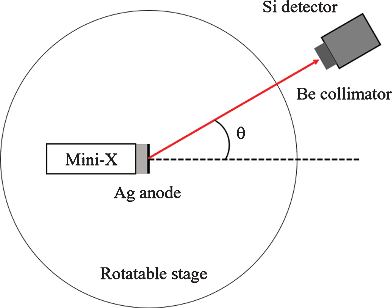
Schematic diagram of the experimental setup.
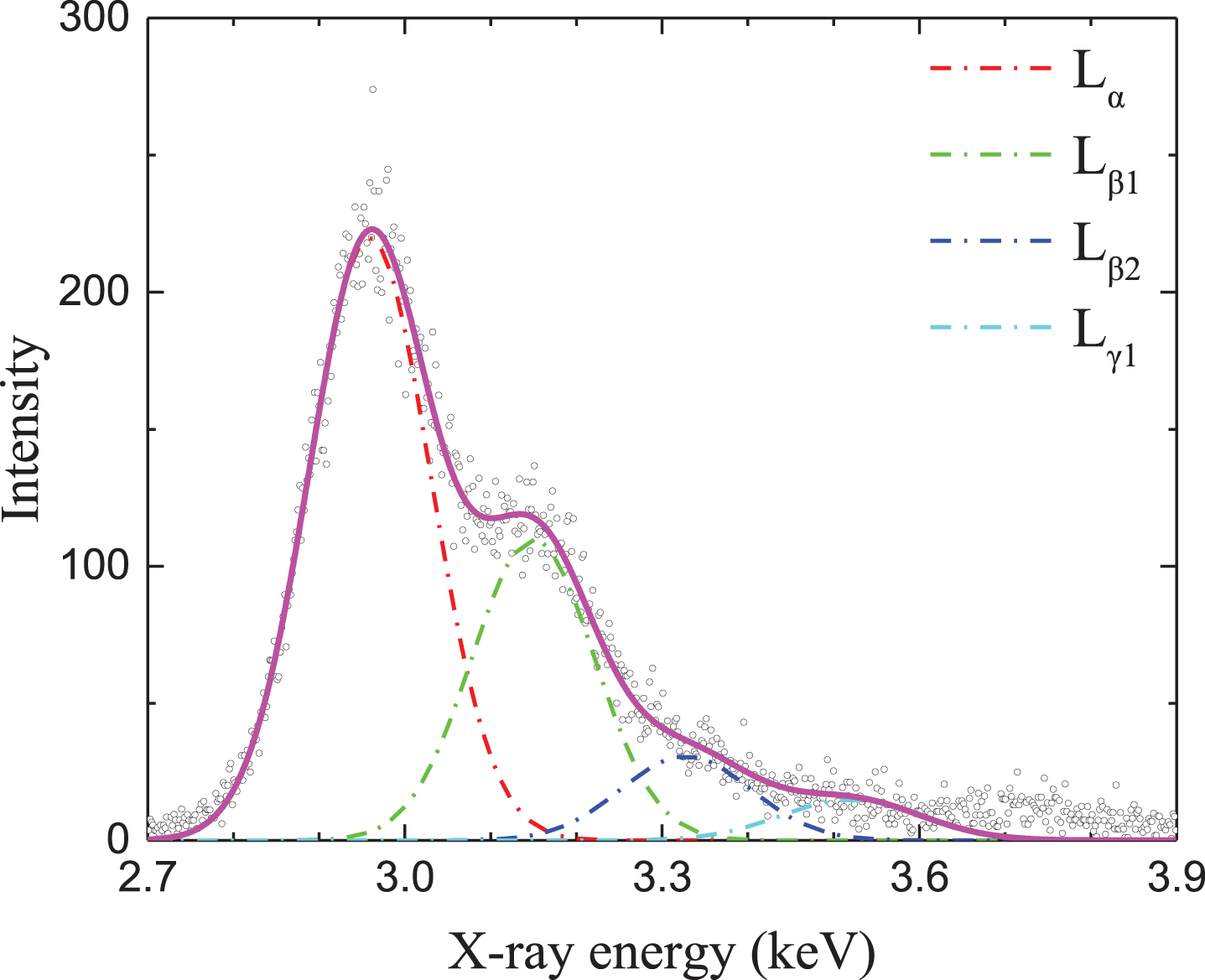
Characteristic L X-ray spectra for Ag at emission angle of 20°. L α , Lβ1, Lβ2 and Lγ1 X-rays are distinguished in the spectra.
The typical spectra for Ag are very similar for detection angle from 0° to 25°. Therefore, it is shown only at one detection angle to introduce the structure of L X-rays for Ag in the paper. The measured characteristic L X-ray spectra at emission angle of 20° are shown in Fig. 2. The energy difference between Ag-Lα1 (L3M5) and Lα2 (L3M4) X-rays is only about 6eV which is much less than energy resolution of X-ray detector. Therefore, both lines cannot be resolved in the measured spectra. Finally, Ag L α (L3M4,5), Lβ1 (L2M4), Lβ2(L3N5) and Lγ1(L2N4) X-rays are distinguished and presented as dashed lines in Fig. 2. The peaks are determined by multi-peak Gaussian fitting procedure. The FWHM of L α , Lβ1, Lβ2 and Lγ1 lines are 168eV, 168eV,179eV and 192eV, respectively, which is consistent with energy resolution of X-ray detector. As a result, net counts for each X-ray lines can be acquired by data processing of the area under corresponding peaks.
In view of Lβ1 and Lγ1 lines originating from 2p1/2 (j = 1/2 ) subshell, they are expected to exhibit isotropic emission. The experimental results actually demonstrate that both lines are found to have isotropic emission spatially within uncertainties in the measurement. Therefore, the angular distribution of Lβ1 and Lγ1 X-ray intensities can be taken as good reference to eliminate the geometry misalignment, if any, in experimental setup. Taking Lβ1 X-ray originating from L2 (2p1/2 , j = 1/2 ) subshell into account, the angular distribution of intensity ratios L
α
/Lβ1, Lβ2/Lβ1 and Lγ1/L
β
1 is investigated in this work. The experimental values of X-ray intensity ratios Lβ2/Lβ1 and Lγ1/L
β
1 can be derived by equation [13]

Intensity ratio of I (L α )/I (L β 1) as a function of P2(cosθ). The dashed line illustrates the trend only.
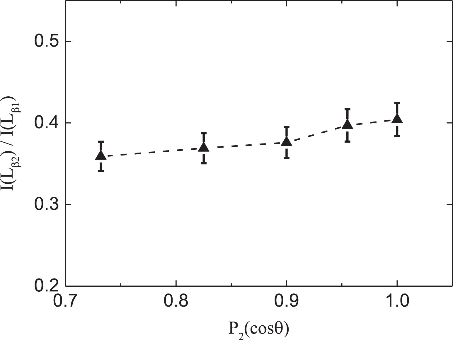
Intensity ratio of I (L β 2)/I (L β 1) as a function of P2(cosθ).
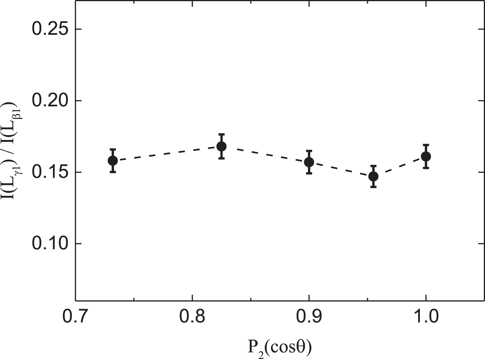
Intensity ratio of I (L γ 1)/I (L β 1) as a function of P2(cosθ).
In electron-impact ionization, the vacancy in L3 subshell can originate not only from direct ionization but also by vacancy transfer from L1- or L2-subshells in Coster-Kronig (CK) process. In addition, the binding energy of electrons in K-shell for Ag (25 keV) is much higher than the energy of incident electrons (15 keV), therefore the electrons in K-shell cannot be ionized and vacancy transfer from K-shell do not need to be taken into account. As a result, the CK correction coefficient κ is expressed by
Theoretical Coster-Kronig yields f ij in L-subshells for Ag taken from Ref. [14]
In the present work, a weighted average value of α= 0.050 is employed according to the relative intensities of Lα1 and Lα2 lines for investigation of alignment degree A20 involving the L α line since both lines cannot be separated with Si(Li) detector. The value of anisotropy coefficient α for Lβ2 line is 0.1, which indicates that the anisotropy parameter β for Lβ2 line is supposed to be twice as much as that for L α line. Consequently, the anisotropy parameter β for Lβ2 lines and the corresponding alignment degree A20 (= β/(ακ) ) for L3-subshell are listed in Table 2. As a result, the alignment property is verified for vacancy in L3-subshell and the alignment degree A20 is derived quantitatively to be 0.65±0.14 in this measurement, while vacancy states in L1- and L2-subshells are not aligned. It should be mentioned that the influence of vacancy transfer from unaligned L1- and L2-subshells can only affect the alignment in L3 subshell slightly.
The measured anisotropy parameter β for Lβ2 line and alignment degree for L3-subshell
As a result, the alignment of L3-subshell is demonstrated and the alignment degree is determined quantitatively. Different from photoionization and ion-impact ionization, we investigate the alignment of atom by electron-impact at 15 keV and consequently the influence of vacancy transfer from K- to L-shell is eliminated since K-shell electrons cannot be ionized. It is also manifested that the CK transition from unaligned L1 and L2 subshells can only affect the alignment in L3-subshell slightly. Comparing to previous work, the alignment degree is determined quantitatively in the experiment.
The angular dependence of L X-ray intensity ratios is investigated for Ag target by 15 keV electron impact. The experimental results suggest that the Lβ2 line has an anisotropic emission spatially; whereas L α , Lβ1 and Lγ1 lines exhibit isotropic emission. Therefore, it is concluded that vacancy states in L1- and L2-subshells are not aligned while vacancy in L3-subshell is aligned. The anisotropy parameters β are determined to be 0.061 for Lβ2 line. The alignment degree A20 is derived quantitatively to be 0.65±0.14 for L3-subshell from the results of anisotropy parameters in this measurement. It demonstrates that the alignment does exist in L3-subshell for atomic ionization by electron impact, and the influence of CK transition from unaligned L1 and L2 subshells can only affect the alignment in L3-subshell slightly. Particularly, the angular correlation effects of X-ray emission originating from L3-subshell will not affect the results significantly in the measurement of L-subshell ionization cross sections on the basis of the current experimental precision.
Footnotes
Acknowledgments
This work is supported by the Natural Science Basic Research Plan in Shaanxi Province of China (Program No. 2018JM1027), and National Natural Science Foundation of China (Grant Nos. 11875219 and 11405123).