
Abstract
Dual-energy computed tomography (DECT) acquires two x-ray projection datasets with different x-ray energy spectra, performs material-specific image reconstruction based on the energy-dependent non-linear integral model, and provides more accurate quantification of attenuation coefficients than single energy spectrum CT. In the diagnostic energy range, x-ray energy-dependent attenuation is mainly caused by photoelectric absorption and Compton scattering. Theoretically, these two physical components of the x-ray attenuation mechanism can be determined from two projection datasets with distinct energy spectra. Practically, the solution of the non-linear integral equation is complicated due to spectral uncertainty, detector sensitivity, and data noise. Conventional multivariable optimization methods are prone to local minima. In this paper, we develop a new method for DECT image reconstruction in the projection domain. This method combines an analytic solution of a polynomial equation and a univariate optimization to solve the polychromatic non-linear integral equation. The polynomial equation of an odd order has a unique real solution with sufficient accuracy for image reconstruction, and the univariate optimization can achieve the global optimal solution, allowing accurate and stable projection decomposition for DECT. Numerical and physical phantom experiments are performed to demonstrate the effectiveness of the method in comparison with the state-of-the-art projection decomposition methods. As a result, the univariate optimization method yields a quality improvement of 15% for image reconstruction and substantial reduction of the computational time, as compared to the multivariable optimization methods.
Keywords
Introduction
Computed tomography (CT) reconstructs a cross-sectional or volumetric image of a patient from a series of projections, providing anatomical and pathological information. Most clinical CT scanner uses a polychromatic x-ray source and x-ray energy-integrating detectors to acquire projection data [1]. CT image reconstruction is typically based on a line integral model, the monochromatic Beer–Lambert law, ignoring x-ray energy information. However, lower energy photons are more easily absorbed than higher energy photons, which causes the x-ray beam hardening effect [2, 3]. Thus, this simplistic line integral model is subject to beam-hardening artifacts in the image reconstruction.
Dual-energy CT (DECT) is a well-established technology to reconstruct material-specific images. Several mainstream technologies include fast kVp modulation (GE Healthcare), dual-source configuration (Siemens Medical Solutions), and dual-layer detection (Philips). The dual layer detectors (Philips’ IQon) consist of two stacked scintillator layers to simultaneously extract low and high energy projection data and have perfect spatial and temporal alignment between low and high energy projections, which is particularly suitable for projection decomposition. According to x-ray imaging physics, in the diagnostic energy range the linear attenuation coefficient mainly reflects the photoelectric effect and Compton scattering. The material-dependent photoelectric and Compton components are functions of material density and atomic number. The photoelectric effect depends strongly on the atomic number and provides the information of material composition, while the Compton scattering is directly proportional to the electron density of the absorber. Based on the energy-dependent non-linear integral model, dual-energy CT can be used to identify the atomic composition and density of material [4, 5], and can be applied for tissue characterization in diagnostic and therapeutic tasks [6].
Several DECT image reconstruction methods were developed over the past years. Alvarez and Macovski simplified the polychromatic non-linear integral equations to two simultaneous cubic equations, which may be solved numerically using a generalized Newton-Raphson method that decomposes the dual-energy measurements into two independent sinograms [4]. A constrained optimization method was also developed to decompose dual-energy projections [7, 8]. Then, statistical methods can be applied to reconstruct images either directly from dual-energy measurements or from material-decomposed sinograms. A joint dual-energy model-based iterative reconstruction (JDE-MBIR) method was proposed to reconstruct basis material densities from the decomposed sinograms [9]. The JDE-MBIR algorithm can be simplified to the polychromatic log-likelihood model using a quadratic approximation to account for noise correlation in material-decomposed sinograms with the constraint of nonnegative x-ray absorption [9]. Basically, these nonlinear optimization methods are computationally expensive and depend on initial values. Hence, the solution obtained using such an optimization method is prone to be trapped at local minima. Alternatively, image-domain material decomposition methods reconstruct images from the low-energy and high-energy sinograms respectively, and then perform material decomposition in the image domain [10, 11]. These image-domain material decomposition methods implicate substantial approximations due to the polychromatic energy spectra and the associated beam hardening effect, resulting in quantitatively inaccurate results [9, 12].
In this paper, a new method for DECT projection decomposition is proposed based on a polychromatic physical model. This approach combines polynomial equation solution and univariate optimization to solve the polychromatic non-linear integral equations in the projection domain. The polynomial equation of an odd order has a unique real solution in the context of DECT, while the univariate optimization is effective to find the global optimal solution. The resultant material decomposition allows accurate and stable DECT image reconstruction. In the next section, the physical model and projection decomposition method are described. In the third section, our numerical simulation and phantom experiments are presented along with representative results. In the last section, relevant issues are discussed.
Methodology
An x-ray source emits a polychromatic x-ray beam, which passes through an object along a path l, and the x-ray intensity I measured by a current-integrating detector can be described by the polychromatic non-linear integral equations according to the Beer-Lambert law [1, 4]:
It is well known that photoelectric absorption and Compton scattering are two dominant x-ray attenuation processes in the diagnostic energy range [4]. The X-ray linear attenuation coefficient is a linear combination of photon-electric effect and Compton scattering [4, 14]. Thus, for compound matter composed of k materials the x-ray linear attenuation coefficient can be expressed as
where
equation 3b is the spatial-dependent photoelectric component for the i-th element, where Z
i
is the atomic number,
equation 3c is the spatial-dependent Compton scattering component,
equation 3d is the energy-dependent photoelectric component [15, 16], where ɛ = E/511 keV , α is the fine-structure constant (≈1/137 ), and r
e
= 2.818 fm is the classical radius of an electron, and
is the energy-dependent Compton scattering component [15], and the Klein-Nishina function [11] is defined as
From Equation (3), we have
where
For projection decomposition, we compute the projections of spatial-dependent photoelectric absorption ∫
l
a (r) dr and the projections of spatial-dependent Compton scattering ∫
l
c (r) dr from DECT measurements I1 and I2. From Equation (5a), we have
where
The fifth-order polynomial approximation to the exponential function is highly accurate for general biomedical imaging. For example, under the following conditions that bone density is 1.92 g/cm3, Z/A ratio of bone 0.51478, muscle density 1.0599 g/cm3, Z/A ratio of muscle 0.55, water density 1.0 g/cm3, Z/A ratio of water is 0.555508, and the x-ray path length of 40 cm through the human body. The error of the fifth-order Taylor approximation will not exceed 0.41% by performing an integral with respect to energy variable ɛ for bone volume fraction of 12.9% or less [17].
By inserting Equation (7) into Equation (6) we obtain that
Equation (8) is a quintic function of a variable x and is strictly convex because the right-hand side of Equation (6) is an exponential function with respect to the variable x. Thus, Equation (8) has a unique real solution in the context of DECT, which is denoted as x = h (y).
Furthermore, inserting function
Therefore, the projection of the spatial-dependent photoelectric absorption can be computed through the following optimization,
Equation (10) can be solved via univariate optimization, such as golden section search and parabolic interpolation. Therefore, the projections of spatial-dependent photoelectric absorption and Compton scattering images can be effectively determined by solving Equations (8) and (10) simultaneously for every detector element at each projection view.
Numerical simulation
A numerical phantom was designed to evaluate the proposed projection decomposition method. The circular phantom has a diameter of 44 cm and contains 9 sub-regions with various materials to simulate human tissues. Each material is composed of several basic elements which are described by the mass density and atomic number, as shown in Table 1. From the composition of the phantom, the photoelectric and Compton scattering images can be accurately computed based on Eqs. (3–4). The x-ray source energy spectral distribution at 140 kVp/30 mA and 80 kVp/30 mA are respectively generated using public software (https://health.siemens.com/booneweb/index.html), as shown in Fig. 1. In the fan-beam imaging geometry, the phantom was placed at the isocenter. The source-to-isocenter distance was set to 62.5 cm, and the source-to-detector distance was 109.7 cm. Along the detector array, there were 828 detector elements equiangularly distributed with 0.109 cm pitch. Based on Eq. (1), the dual-energy projection datasets were generated with 360 views over a range of 360°. Then, the projection data were corrupted by Poisson noise to simulate real experimental conditions. From the DECT projection datasets, the proposed univariate optimization method was applied to estimate the projection components of spatial-dependent photoelectric absorption and Compton scattering respectively. The calculated results are shown in Fig. 2.
Physical parameters of each component in the numerical phantom [18]
Physical parameters of each component in the numerical phantom [18]
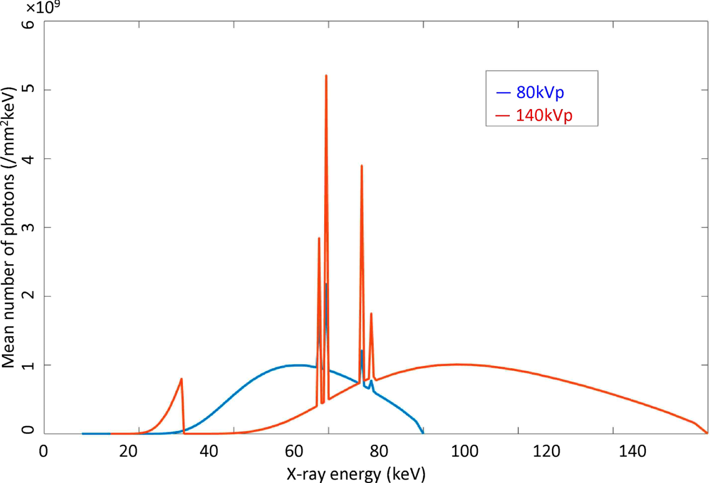
Energy spectral distributions of the x-ray source. (a) The energy spectrum of the x-ray tube operated at 80 kVp and (b) the energy spectrum of the x-ray tube at 140 kVp.
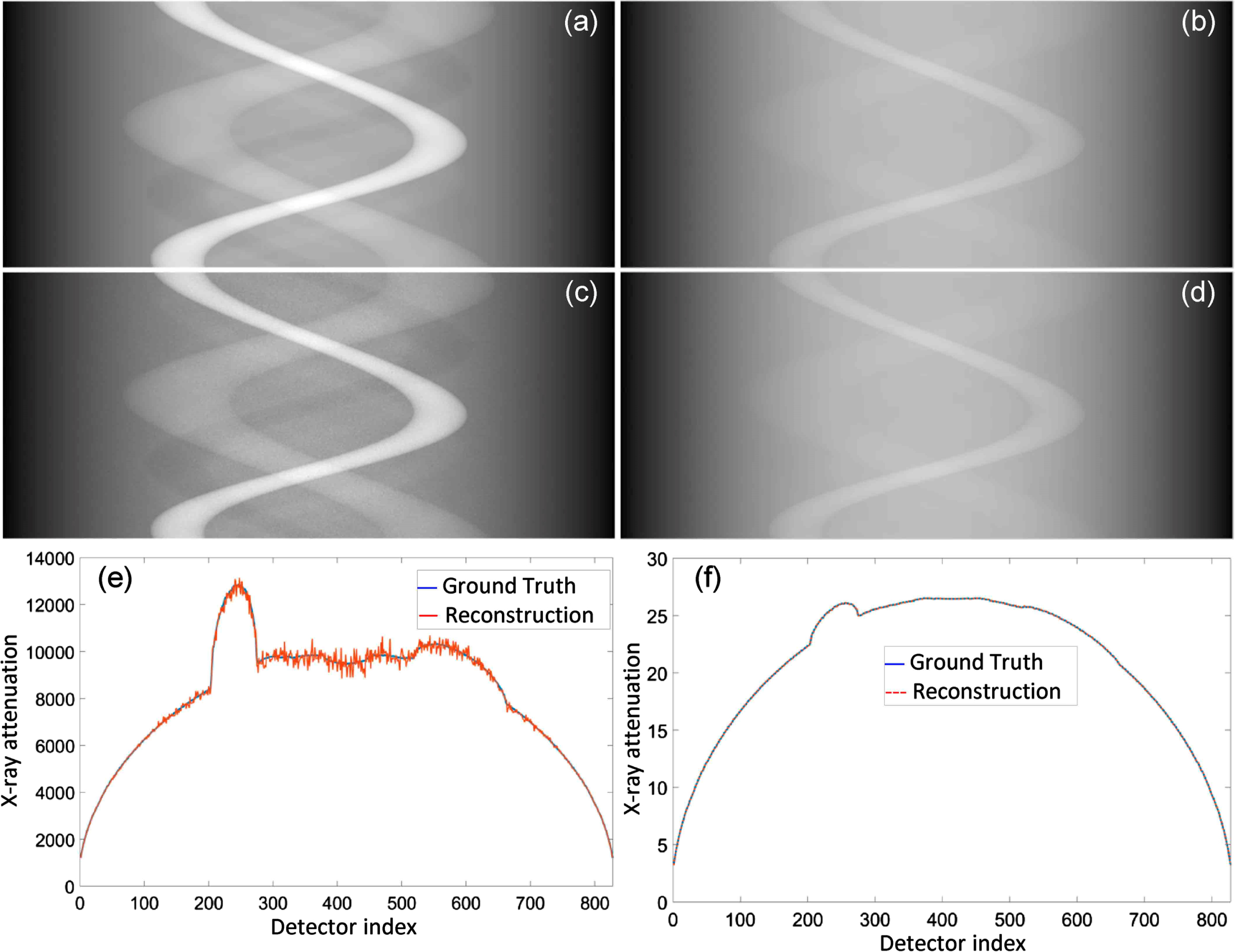
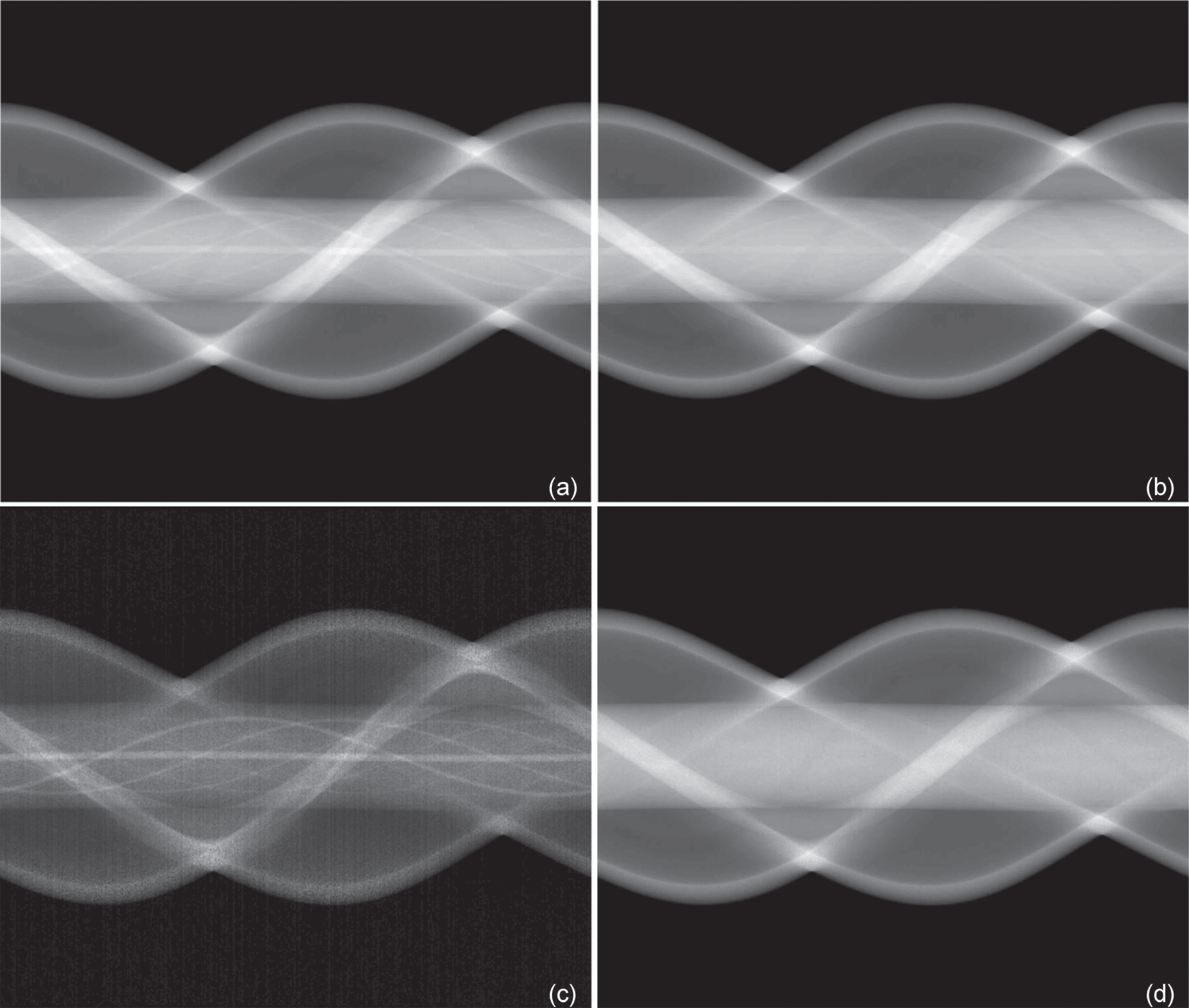
Projection decomposition of spatial-dependent photoelectric absorption and Compton scattering. (a) and (b) are the ground-truth sinograms of spatial-dependent photoelectric absorption and Compton scattering respectively; (c) and (d) the decomposed sinograms of spatial-dependent photoelectric absorption and Compton scattering using the univariate optimization method; (e) and (f) the representative ground-truth and reconstructed profiles in the first imaging view of photoelectric absorption and Compton scattering respectively.
We also performed the projection decomposition using the multivariable optimization method to reconstruct photoelectric absorption and Compton scattering projections for comparison [7]. In the numerical simulation, we found that the multivariable optimization method sometimes converged prematurely to an inaccurate solution if the initial guess is not close enough to the true solution. For our numerical test, we used the estimated solution of linearization equations (5a)–(5b) as the intial values. The projection decomposition results are shown in Fig. 3.
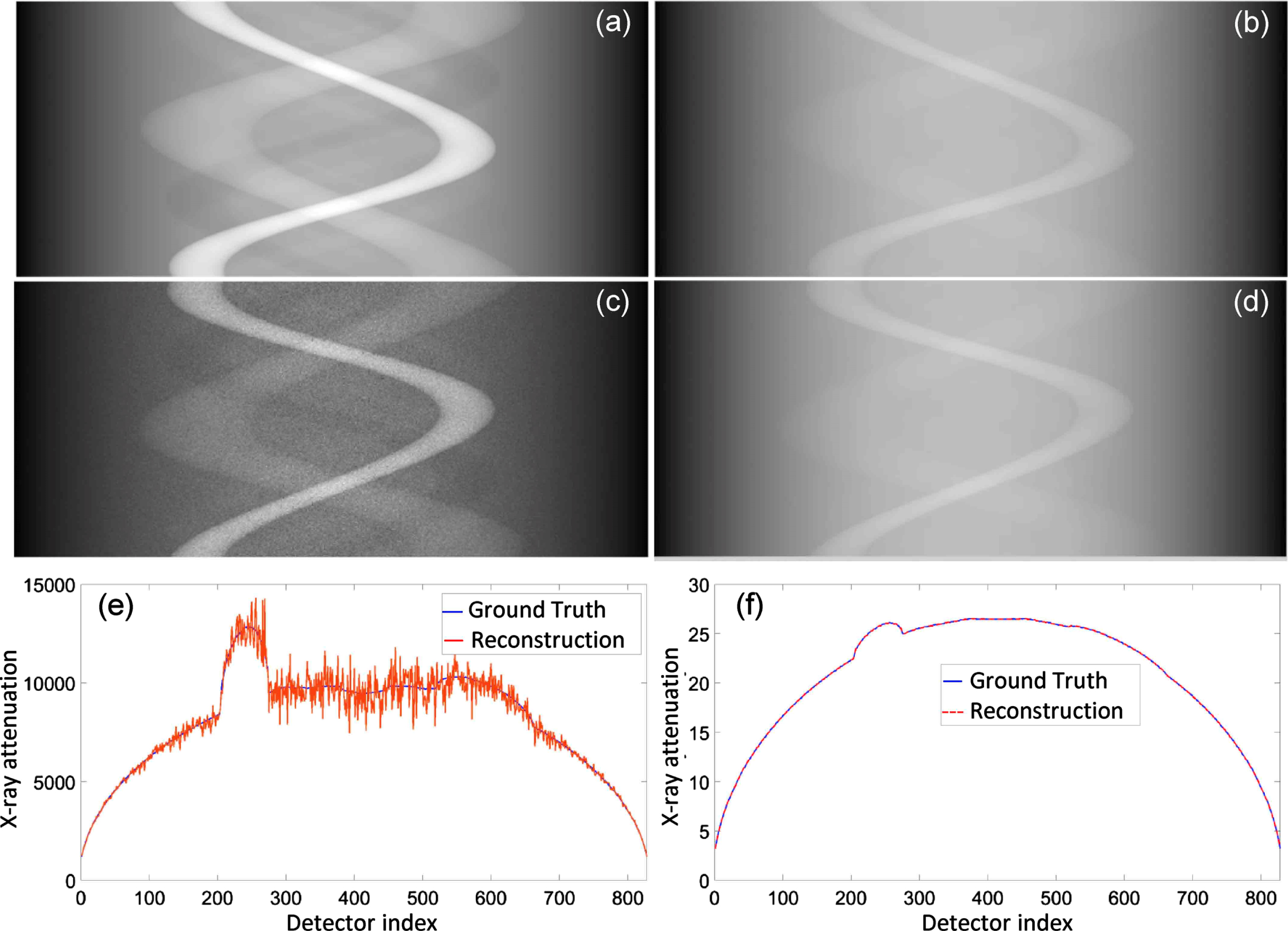
Comparative study against the multivariable optimization method. (a) and (b) are the ground-truth sinograms of spatial-dependent photoelectric absorption and Compton scattering respectively; (c) and (d) the sinograms of spatial-dependent photoelectric absorption and Compton scattering images reconstructed using the multivariable optimization method; (e) and (f) the ground-truth and reconstructed profiles in the first projection view of photoelectric absorption and Compton scattering projections respectively.
From the reconstructed sinograms of photoelectric absorption and Compton scattering, image reconstructions were performed using the simultaneous algebraic reconstruction technique (SART). The reconstructed photoelectric absorption and Compton scattering images using our univariate optimization method were presented in Fig. 4(c–d), showing decent spatial and contrast resolution, and texture features. The reconstructed photoelectric absorption and Compton scattering images using the multivariable optimization method were shown in Fig. 4(e–f).
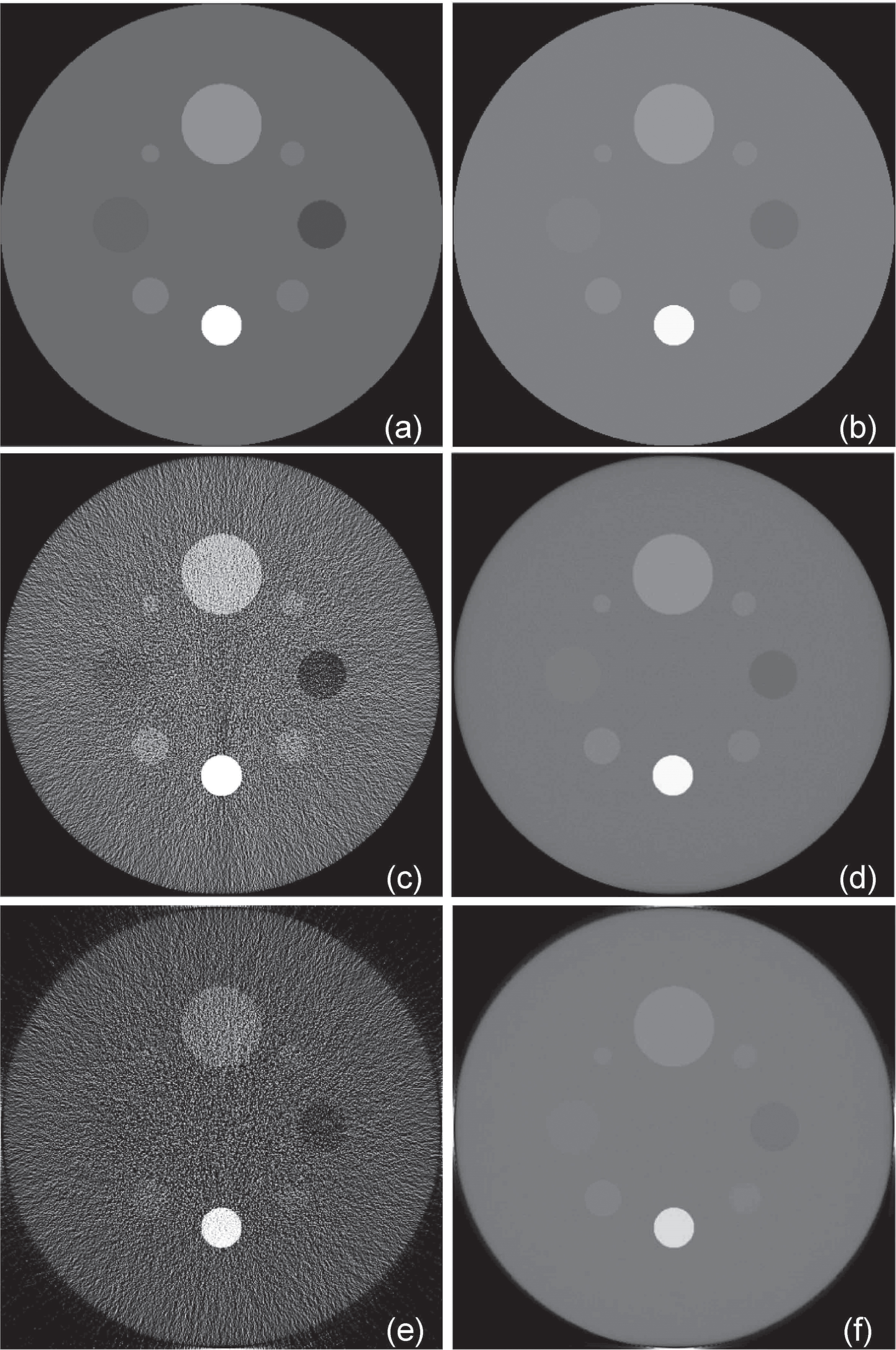
Images in terms of photoelectric absorption and Compton scattering respectively. (a) and (b) are the ground truth photoelectric absorption and Compton scattering images respectively, (c) and (d) the photoelectric absorption and Compton scattering images reconstructed using the univariate optimization method, (e) and (f) the photoelectric absorption and Compton scattering images reconstructed using the multivariable optimization method.
Furthermore, the reconstructed images were evaluated in terms of the peak-signal-to-noise ratio (PSNR) and the structural similarity index measure (SSIM). The photoelectric absorption images reconstructed using our univariate optimization method achieved an average PSNR of 23.21 and SSIM of 0.686, while the counterparts reconstructed using the multivariable optimization method achieved an average PSNR of 15.31 and SSIM of 0.455. The Compton scattering images reconstructed using the univariate optimization method achieved an average PSNR of 26.73 and SSIM of 0.949, while the counterparts reconstructed using the multivariable optimization method achieved an average PSNR of 17.42 and SSIM of 0.869. The univariate optimization yields a quality improvement of 15% for the image reconstruction at a substantially reduced computational cost (about 10%) comparing to the multivariable optimization method. From the reconstructed Compton scattering and photoelectric absorption images, virtual monochromatic images can be calculated based on Equation (4). Accordingly, the effective atomic number (EAN) images and electron density (ED) images can be also obtained, which are valuable for many clinical applications such as proton therapy.
Experiments with actual DECT scans of a customized physical phantom were conducted to evaluate the proposed reconstruction method. The phantom consists of vessel inserts in a homogeneous tissue-equivalent cylindrical background, which was then inserted in a QRM pseudo-anthropomorphic thorax phantom, as shown in Fig. 5. The phantom contains twelve cylindrical holes of diameters 1.0, 1.1, 1.2, 1.3, 1.4, 1.5, 1.7, 2.0, 2.5, 3.0, 4.0, 5.0 mm respectively, which were filled with 30-mg/mL iodine-water solution. The thorax phantom was scanned by a 256-slice GE Revolution CT scanner. An axial scan was performed to generate 984 projections over a 360 degree angular range. Only the data from the center detector rows were used in this study. Low-energy/high-energy projection datasets were acquired by setting x-ray tube at 80 kVp/50 mA and 140 kVp/35 mA respectively, with 1 second/rotation. We obtained a set of fan-beam sinograms at 80 kVp and 140 kVp respectively, as shown in Fig. 6(a–b).

DECT reconstruction through a middle slice of the QRM phantom.
Sinograms of a thorax phantom acquired using the GE Revolution CT scanner with (a) 80 kVp/50 mA and (b) 140 kVp/35 mA respectively. (c) and (d) are the sinograms of spatial-dependent photoelectric absorption image and Compton scattering image reconstructed using the univariate optimization method.
Clearly, photoelectric absorption and Compton scattering modeling is equivalent to two-material decomposition [19]. In practice, it is usually convenient to represent the energy-dependent attenuation as a linear combination of basis materials. Each pixel is comprised of a linear combination of pre-selected target materials (e.g., iodine and water). The material-based decomposition algorithm generates the two material specific images. Hence, the mass attenuation coefficient of mixtures can be computed as a weighted average of the mass attenuation coefficients of basis materials according to the additivity principle [15, 20]:
where
For the QRM phantom, we performed material-specific image reconstruction. In this case, the univariate optimization algorithm described in Section 2 still works for matter-based decomposition by Equation (12). From the measured sinograms at the low (80 kVp/50 mA) and high (140 kVp/35 mA) kVp settings, projection data were decomposed into the sinograms of two basis materials, as shown in Fig. 6(c–d). Furthermore, the water and iodine basis images were reconstructed from the decomposed sinograms using the SART algorithm, as shown in Fig. 7. It is observed in Fig. 7 that the material decomposition was achieved accurately using our univariable optimizaton method. By calculating average pixel-wise errors within the cylindrical holes in the iodine basis image, the average mass fraction of the iodine solution was 2.5%, yielding a quantitative error of about 17%.
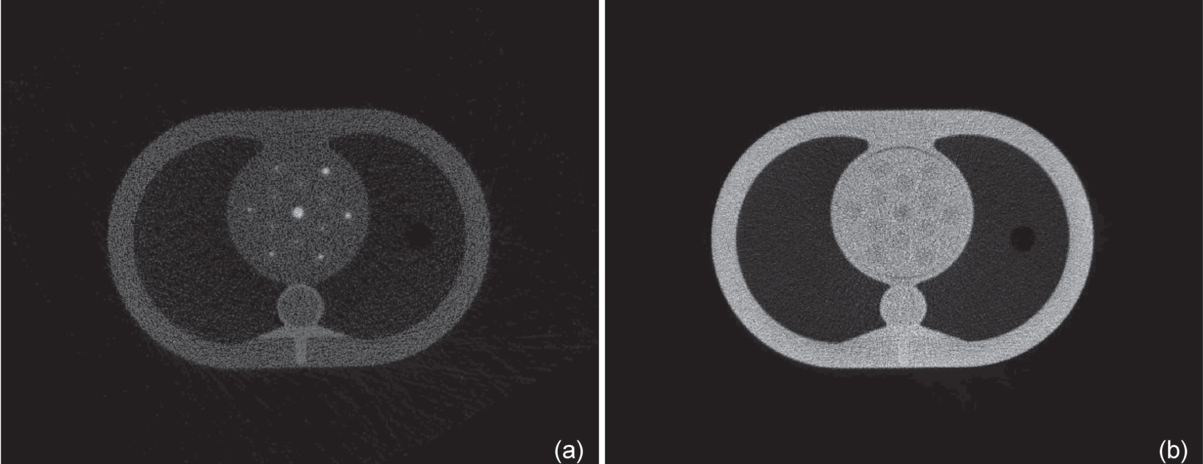
Material decomposition images. (a) The Iodine and (b) water basis images reconstructed from the reconstructed material basis sinogram.
In this study, a novel projection decomposition method has been proposed to solve the system of polychromatic non-linear integral equations from two projection datasets acquired at low and high x-ray energy spectra respectively. This method combines polynomial modeling and univariate optimization to perform DECT projection decomposition. Because the polynomial function is strictly convex, the polynomial equation of an odd order has a unique real solution in the context of DECT, and univariate optimization can be easily performed to find the global optimal solution. Our numerical and physical experiments have shown that the proposed image reconstruction method allows more accurate and stable projection decomposition compared to the state-of-the-art multivariate optimizaiton method. As a result, the univariate optimization method yields a quality improvement of 15% for image reconstruction and also about 10% reduction of the computational cost.
Material decomposition has been performed to identify specific materials according to their effective atomic number and mass density. In the diagnostic x-ray energy range, the mass attenuation coefficient depends only on the chemical composition and effective x-ray energy. Based on the energy-dependent photoelectric–Compton model, the virtual monochromatic images (VMI) can be obtained. The effective atomic number (EAN) images and electron density (ED) images can be also used for various applications such as planning for proton therapy [21].
Further extensions of the work will incorporate deep learning to enhance the quality of projection decomposition. Emerging deep learning techniques can extract non-linear relationships, discover complicated features and representations in a data driven manner. Deep neural networks can help improve various types of diagnostic and interventional tasks. These have been successfully applied for super-resolution imaging [22], image denoising [23, 24], and virtual monoenergetic imaging [20]. Clearly, deep learning techniques are valuable for denoising and deblurring of photoelectric absorption and Compton scattering images. A number of interesting topics on material decomposition can be also investigated in the future.
Footnotes
Acknowledgments
This work is partially supported by the National Institutes of Health Grants NIH/NIBIB R01CA237267 and R01EB026646.