
Abstract
Objective:
The objective of this mini-review is to examine a subset of literature that demonstrates multiple interactions between mechanics and biology within the spine and propose how incorporation of these mechano-biologic interactions can be applied to improve the conceptual understanding of tissue tolerances.
Background:
Low back pain represents a major musculoskeletal problem in the workplace. Traditional biomechanical assessments have employed tissue tolerances as an approach for reducing workplace injuries; however, development of more universal biologically sensitive tolerances requires incorporation of mechano-biologic interactions.
Methods:
A focused literature review addressing the interactions between mechanical loading and biology in the spine.
Results:
Mechanical loads applied to the body are distributed across all spatial scales from the body to the tissues to the cells. These mechanical loads regulate cellular metabolism and over time can lead to tissue strengthening or weakening. Mechanical loading also interacts with the biologic environment (e.g., tissue inflammation, nerve sensitization) to influence the perception of pain, thereby changing the risk of experiencing pain. Biologic tissues also exhibit time-dependent changes in mechanical behaviors that occur throughout the day and with disease, suggesting tissue tolerances are time dependent.
Conclusion:
Incorporating mechano-biologic interactions into the traditional tissue tolerance paradigm through describing tissue tolerances as a function of multiple factors (e.g., preexisting risk factors, underlying pathology, and time) may lead to the development of tissue tolerances that are more representative of the in vivo situation.
Application:
Efforts must work toward incorporating biological concepts into tissue tolerances in order to improve risk assessment tools.
Introduction
Chronic low back pain (LBP) is the leading cause of disability worldwide, with immense socioeconomic costs (>$100 billion in the United States alone) (Katz, 2006; Vos et al., 2012). In particular, disorders of the intervertebral disc (IVD) are commonly thought to contribute to the development of low back pain (Freemont, 2009; Freemont et al., 1997) and are the target tissue for a large proportion of clinical treatments. A primary goal of ergonomics research has been to prevent or control the occurrence of low back pain in the workplace. A traditional ergonomic paradigm compares the loads the tissue experiences to a reference load, or tissue tolerance, which is the load limit above which an individual is at a greater risk of injury or pain. Although this ergonomic paradigm is a valuable tool, it does not incorporate the tissue’s biologic response to loading, which includes changes in structure with disease and the tissue’s ability to actively respond to its mechanical environment. Therefore, inclusion of the tissue’s dynamic biologic response to mechanical loading may enable the development of more universal tissue tolerances in living people. The objective of this mini-review is to highlight a subset of the literature that demonstrates different interactions between mechanics and biology and suggest how incorporation of these interactions can be applied to improve the conceptual understanding of tissue tolerances and to prevent injury.
The Premise
The spine functions through an intricate combination of biomechanical and biologic factors that interact in different spatial and temporal scales. A key concept underlying the interaction between mechanics and biology is that the cells within the tissue respond to a mechanical input and that this cellular output can modify the mechanical response of the tissue. When mechanical forces are applied to the body, the forces are distributed across all spatial scales, that is, from the whole body, to the spine, to the individual tissues, and eventually down to the cells (Figure 1). Inherent in this multiscale concept is that biologic tissues are composed of two primary components: the extracellular matrix, which is all of the noncellular components of the tissues, and the cells embedded within the extracellular matrix. The composition, physical properties, and alignment/orientation of the noncellular components within extracellular matrix are what give the tissue most of its mechanical integrity. As such, any changes to the extracellular matrix, which may result from genetic variation, disease, mechanical damage, or tissue synthesis/degradation, will have a direct effect on the ability of the tissue to bear load and thereby influence its mechanical tolerance.
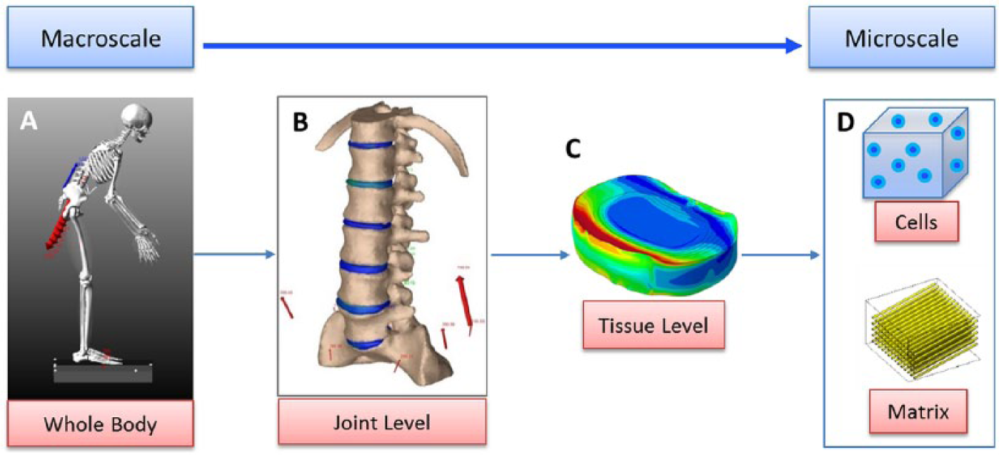
Multiscale spinal mechanics. Mechanical forces are distributed across all spatial scales, that is, from the whole body to the spine to the individual tissues and eventually down to the cells. (A) Loads that are applied to the whole body are distributed to the (B) spinal motion segments and then transferred to (C) the individual spinal tissues and down to (D) the extracellular matrix and sensed by the embedded cells.
Current tissue tolerance limits within the lumbar spine have been based primarily on the onset of mechanically induced tissue damage (Marras, 2012). Historically, the tissue tolerance for compressive forces within the lumbar spine (forces acting down the long axis of the spine) have been based on the onset of endplate microfracture (Chaffin, Andersson, & Martin, 2006). However, recent work has also started to incorporate other types of loading, such as shear loading (forces acting perpendicular to the compressive forces), and also to incorporate the interaction between the number of repetitions of applied loading and the loading magnitude (Gallagher & Marras, 2012). All of these parameters are important, especially in work-related scenarios that include repetitive tasks. We propose that in addition to the factors listed previously, incorporation of changes that occur to the extracellular matrix with degeneration and in response to biologic factors will allow the development of potentially more universal tolerance limits. This mini-review highlights the interactions between biomechanical loading and intervertebral disc biology and focuses on how an improved understanding of this complex interplay might provide insights into the development of new tolerance limits in the future.
Multifactorial Etiology of Low Back Pain
Epidemiological studies have demonstrated that many factors contribute to the risk of developing low back pain (Battie, Videman, Levalahti, Gill, & Kaprio, 2007; Linton, 2000; Norman et al., 1998; Steffens et al., 2015). The plethora of risk factors can be grouped into three broad classifications: biological/individual (e.g., genetics, gender, age, anthropometry, alcohol/smoking, back pain history), physical (e.g., heavy physical work, static sedentary work, lifting/forceful movements, whole body vibration, awkward postures), and psychosocial (e.g., high demand jobs, high perceived stress, job dissatisfaction) (Battie et al., 2007; Linton, 2000; Norman et al., 1998; Steffens et al., 2015). Despite the identification of multiple important epidemiological factors, uncertainty remains in our understanding of the causal factors of low back pain as many of these factors can vary based on the degree, or dosage, of exposure. Many of these risk factors also likely interact at various levels of intensity, and this interaction influences overall risk. For example, the relevance of a genetic defect affecting extracellular matrix integrity may increase as that individual is exposed to higher physical loads. These interactions are not adequately chronicled by epidemiological studies, and so only a partial view of the complex problem is provided (Marras, 2008).
Mechanical Loading Influences Matrix Composition
The spine experiences millions of loading cycles over decades of life. These loading cycles play an important role in regulating the mechanical integrity of the tissue, similar to how you can build or lose muscle through exercise or a sedentary life style. The cells within the tissue perceive and respond to mechanical loading by changing their metabolic balance between matrix synthesis (tissue build-up/production) and matrix breakdown (Figure 2). Mechanical loading is well recognized to regulate the structure and function of musculoskeletal tissues ranging from bone, muscle, cartilage, and the IVD (Bader, Salter, & Chowdhury, 2011; Killian, Cavinatto, Galatz, & Thomopoulos, 2012; Neidlinger-Wilke et al., 2014; Sugiyama, Price, & Lanyon, 2010). When mechanical loading is applied to a tissue, the mechanical response of the tissue is dependent on the amount of tissue (cross-sectional area) that the force is acting on, similar to how a thin piece of rope may break under a given tensile load and a thick piece of rope would not break under the same load. Therefore, when mechanical loading is applied to tissues, it is generally described as a pressure, or mechanical stress, and is expressed in units of force (N) per cross-sectional (m2) area or Pascal (Pa). Specifically in the healthy IVD, dynamic loading within a “physiologic” range of pressures and frequencies (0.2–1 MPa at 0.1–1.5Hz) is largely beneficial and shifts the metabolic balance to favor matrix synthesis while reducing matrix breakdown (Chan, Ferguson, & Gantenbein-Ritter, 2011; Korecki, MacLean, & Iatridis, 2008; MacLean et al., 2003; MacLean, Lee, Alini, & Iatridis, 2004, 2005). However, if external loading is either above or below this physiologic range, the cellular response is no longer beneficial and shifts toward favoring matrix breakdown (Chan et al., 2011; Paul et al., 2013; Stokes & Iatridis, 2004).

Mechanical loading influences matrix composition and injury risk. The biologic response of a tissue is always a balance between tissue build-up and breakdown. (A) The mechanical integrity of the tissue is maintained when there is equal amounts of tissue build-up and breakdown. (B) The tissue is weakened when tissue breakdown is greater than the tissue build-up, and (C) the tissue is strengthened when tissue build-up is greater than breakdown. (D) The conceptual relationship between loading and cellular metabolism demonstrating that the metabolic balance is influenced by the type of loading the tissues experience, which can change the balance between matrix breakdown and matrix build-up/synthesis. (E) The risk of injury (R) at a given physical work intensity is influenced by the accumulation of matrix turnover and can be reduced in strengthened tissues (Rstrong) or increased in weakened tissues (Rweak).
Adaptation of the extracellular matrix to mechanical loading occurs over a long temporal scale and suggests that tissue integrity, or tissue quality, is dynamic and can be strengthened and weakened over time. This dynamic tissue quality likely contributes to the high variability in tissue tolerances previously described (compression: 3 kN–8 kN) (Marras, 2012) and may also help explain the variability in the relative risk of experiencing low back pain at a given physical work load exposure. For example, a weakened tissue may have an elevated risk of injury while a strengthened tissue may have a reduced risk of injury compared to a normal tissue.
Biomechanics, Biology, and Pain
Another example of how biomechanics and biology interact is in pain perception. It is well established that pain originates from pain sensing nerves (i.e., nociceptors). However, induction of pain can arise from multiple sources. A common source of low back pain involves impingement of the spinal cord or a nerve root following an IVD injury or herniation. Although these types of injuries are prevalent among clinical populations, pain can also arise from degeneration of other spinal tissues such as ligaments, facets, or the IVD.
The healthy IVD is the largest organ in the body that does not have a direct peripheral nerve or blood supply; however, with disease progression, nerves and blood vessels have been demonstrated to grow into painful discs through the endplate and outer region of the IVD into the inner regions (Fields, Liebenberg, & Lotz, 2014; Freemont et al., 2002). There is a limited understanding of the mechanisms driving this aberrant nerve growth into the IVD; however, changes in the mechanical integrity and biochemical environment of the IVD tissue have been suggested as potential mechanisms promoting nerve growth (Stefanakis et al., 2012; Stefanakis, Luo, Pollintine, Dolan, & Adams, 2014). Matrix breakdown that occurs following injury or with aging and degeneration can lead to a loss of proteoglycans, a key component of the extracellular matrix within the IVD. Proteoglycans play an important role in preventing nerve and blood vessel growth (angiogenesis) through (a) directly inhibiting nerve growth (Johnson, Caterson, Eisenstein, & Roberts, 2005; Johnson et al., 2002; Purmessur et al., 2015) and (b) inducing a hydrostatic swelling pressure within the IVD that physically prevents nerve growth. Therefore, a loss of proteoglycans leads to a loss of the direct inhibition as well as reductions in the intradiscal pressurization, thereby creating an environment that is both mechanically and biochemically conducive for nerve and blood vessel growth (Stefanakis et al., 2012). A second contributing factor is the presence of biochemical signals that promote/direct neuronal growth and angiogenesis, such as pro-inflammatory signals and growth factors (Abe et al., 2007; Purmessur, Freemont, & Hoyland, 2008; Shafiq, Jung, & Kim, 2015). These factors can be expressed by IVD cells and by the surrounding blood vessels, which can penetrate the IVD once structural disruption has occurred, and direct nerve growth.
Although the presence of nerves is necessary for the sensation of pain, some form of nerve stimulation is also required for a pain response to be induced. Mechanical forces acting on nociceptors can induce pain; however, it is also known that biochemical stimulation of nociceptors (i.e., inflammatory proteins such as pro-inflammatory cytokines) can also induce a pain response (Garcia-Cosamalon et al., 2010; Schaible, 2014). In vivo, these biochemical and mechanical factors likely interact to influence pain perception. It is understood that an individual’s pain threshold may be different than the tissue tolerance limit, especially in poorly innervated tissues such as the IVD. In such tissues, changes in the biochemical environment (i.e., tissue inflammation) may provide two potential mechanisms for elevated sensitivity to pain. The first mechanism of elevated sensitivity is that the mere presence of certain biochemical factors, such as pro-inflammatory cytokines, can “sensitize” local nociceptors, meaning that the neuronal activation threshold is lowered so that typically innocuous stimuli can induce a pain response (Izzo, Popolizio, D’Aprile, & Muto, 2015; Schaible, 2014). Thus, typically nonpainful loading may induce a pain response in nerves that have been sensitized (Figure 3). The second consequence of inflamed tissue is that local swelling may occur that increases the hydrostatic pressure on nerves, potentially causing pain or at least reducing the amount of additional external loads required to elicit a pain response.
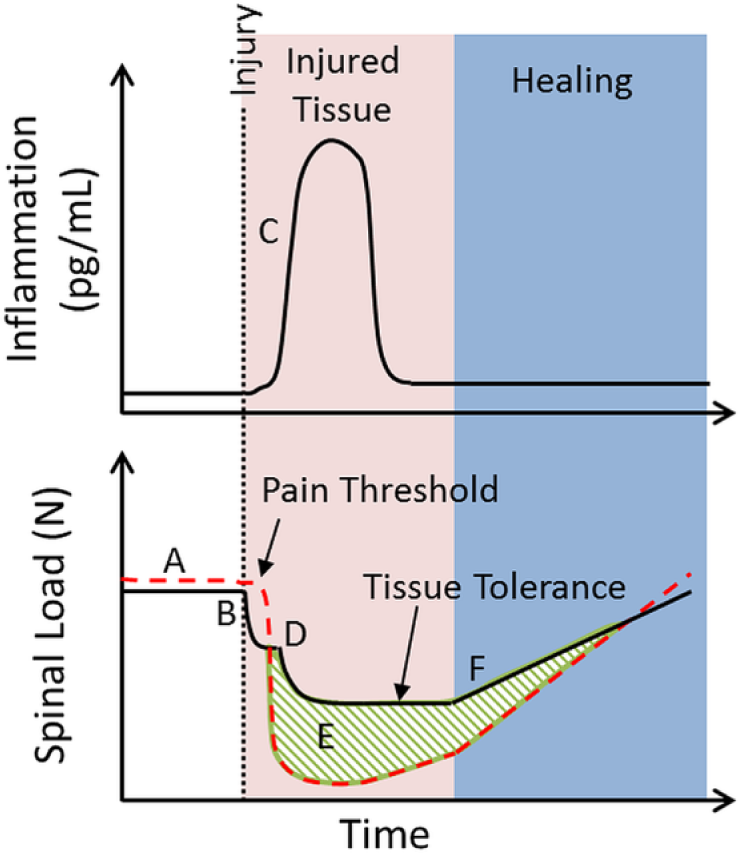
Conceptual relationship between pain sensitivity, inflammation, and load tolerance following tissue injury. (A) Initially, there is minimal tissue inflammation, and the tissue tolerance, the load limit above which an individual is at a greater risk of injury, is below the pain threshold due to the limited innervation within the intervertebral disc (IVD). (B) Tissue injury occurs, and there is an instantaneous decrease in the tissue tolerance. (C) There is an increase in tissue inflammation following tissue injury. (D) The presence of inflammation induces matrix breakdown, further decreasing tolerance, and sensitizes the nerves, resulting in a drop in the pain threshold. (E) As a result of the drop in the pain threshold, normal loading that is not damaging and is below the tissue tolerance can still induce pain. (F) Gradual resolution of inflammation and tissue healing/strengthening occurs.
Mechanical loading itself can also contribute to the presence of pro-inflammatory mediators within the IVD, through (a) facilitating the transport of pro-inflammatory cytokines into the IVD from the surrounding tissues (Walter et al., 2015) and (b) inducing the expression of pro-inflammatory cytokines by the native IVD cells (Gawri et al., 2014; Walter et al., 2011). The presence of multiple pro-inflammatory cytokines is associated with matrix breakdown seen in IVD degeneration and is positively correlated with aging and severity of degeneration (Bachmeier et al., 2007; Le Maitre, Hoyland, & Freemont, 2007; Risbud & Shapiro, 2014; Weiler, Nerlich, Bachmeier, & Boos, 2005). Cell culture experiments have demonstrated that these pro-inflammatory cytokines induce a metabolic shift favoring matrix breakdown (Hoyland, Le Maitre, & Freemont, 2008; Le Maitre et al., 2007; Purmessur et al., 2013; Seguin, Bojarski, Pilliar, Roughley, & Kandel, 2006; Seguin, Pilliar, Roughley, & Kandel, 2005), and organ culture experiments have confirmed that a prolonged exposure to inflammatory mediators can directly alter the mechanical behavior of the IVD (Walter et al., 2015). Overall, these studies highlight that the mechanical and biochemical environments can mediate nerve growth and influence nerve stimulation and that these events likely coincide with changes in the local loading environment within the IVD following matrix breakdown.
Time-Dependent Mechanical Behaviors
Biological tissues are viscoelastic and exhibit time-dependent changes in mechanical behavior. The tolerance of a tissue is dependent on its mechanical properties; however, the mechanical properties of a tissue can change with time and suggest that the tolerance of that tissue may also be time dependent. In the spine, this time-dependent mechanical behavior is most prominently observed in the IVD and is a result of changes in tissue hydration that occur over time (Adams, Dolan, & Hutton, 1987; Urban & McMullin, 1988). The IVD’s water content can vary by up to 20% throughout the day (Botsford, Esses, & Ogilvie-Harris, 1994) and also progressively decreases as degeneration progresses. This results in the IVD experiencing changes in mechanical behaviors on two time scales: one that occurs on a daily basis as the IVD compresses, or creeps, throughout the day and a second that occurs over decades as the extracellular matrix is degraded and broken down during aging and degeneration (Figure 4).
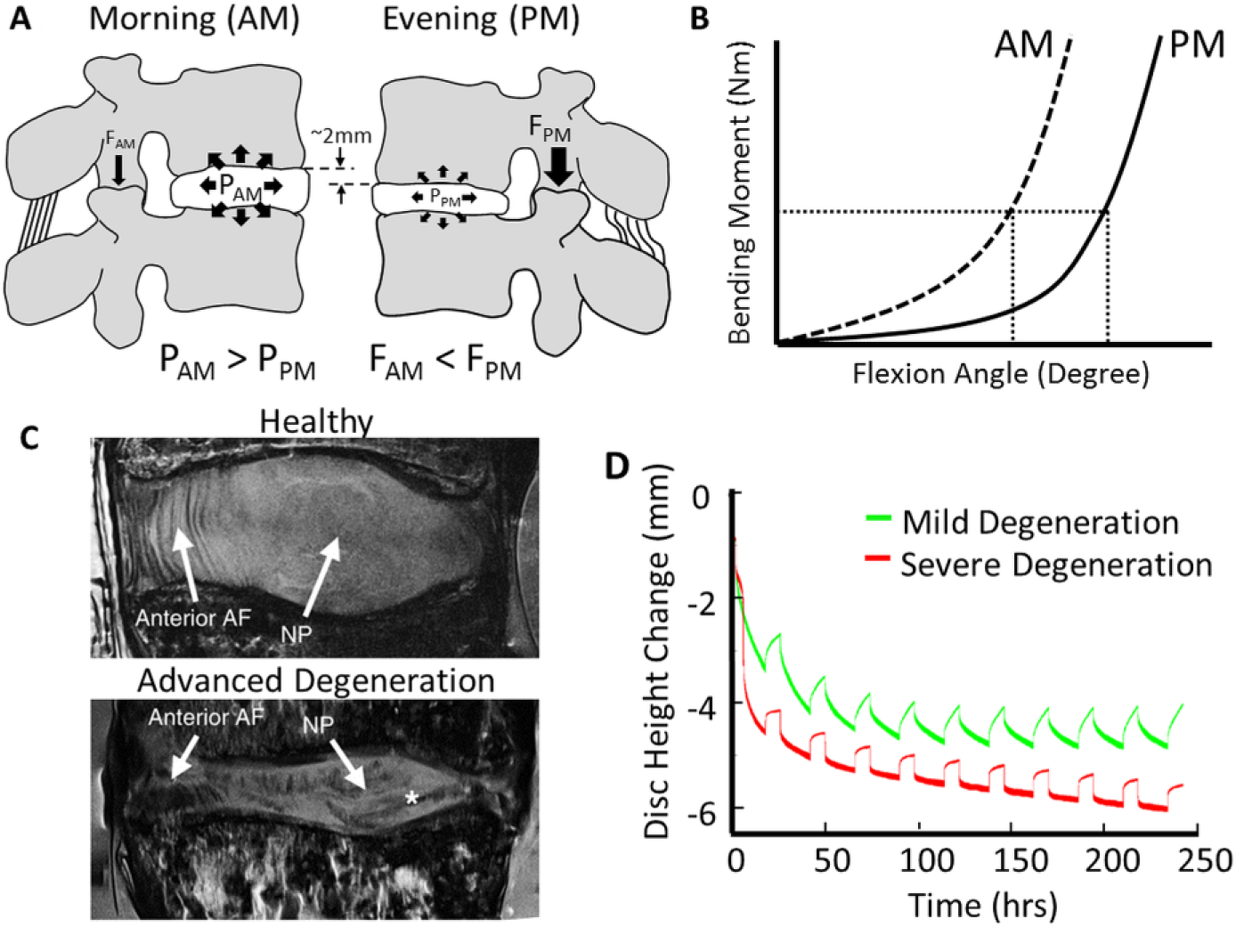
Time-dependent mechanical behavior of the intervertebral disc (IVD). The spine experiences time-dependent changes in mechanical behavior that occur on two time scales: daily and over decades. (A) Schematic demonstrating the diurnal change in IVD height and the resultant change in ligament laxity, facet contact forces (F), and nucleus pulposus (NP) pressurization (P) in the morning (AM) and the evening (PM). (B) Schematic demonstrating the diurnal change in height influences the mechanical behavior of the spine, which is evident by an increased degree of motion segment flexibility. (C) Sagittal magnetic resonance imaging of human lumbar IVDs demonstrating the changes in structure that occur within the annulus fibrosus (AF) and NP regions of the IVD during degeneration. Images modified from Smith, Nerurkar, Choi, Harfe, and Elliott (2011). (D) Examples of the differences in diurnal height loss (24-hour cycle) in human lumbar IVDs of mild and severe degeneration cultured under the same loading conditions (16 hours: 370 ± 130 kPa, 8 hours: 73 ± 10 kPa) in organ culture. Figure modified from Emanuel et al. (2015) with permission.
The IVD experiences an 8% to 10% height loss throughout the day with a total change in the length of the spine of ~19 mm between the morning and the evening (Tyrrell, Reilly, & Troup, 1985; Walter, Illien-Junger, Nasser, Hecht, & Iatridis, 2014). This daily disc height loss increases the compressive stiffness of the IVD and reduces intradiscal pressures as water is lost, leading to a redistribution of the spinal loads primarily on the IVD in the morning to other spinal structures in the evening (Adams, Dolan, Hutton, & Porter, 1990; Zander, Krishnakanth, Bergmann, & Rohlmann, 2010) (Figure 4A). This loss of disc height also results in an overall increase in spinal flexibility and range of motion and can be thought of as a decrease in the spine’s resistance to bending. This decreased resistance to bending is evident in that the flexion angle of the spine increases under the same bending moment, which is a measure of the tendency of a structure to bend from an applied force (Adams & Dolan, 1996; Jamison & Marcolongo, 2014; Zander et al., 2010) (Figure 4B). These diurnal changes in IVD mechanics are thought to be clinically significant and may contribute to the elevated odds ratios for the onset of acute low back pain in the morning, when compressive loading induces greater pressurization within the IVD, versus the afternoon (Adams et al., 1990; Steffens et al., 2015).
The composition of the extracellular matrix within the IVD changes throughout the degeneration process and is progressively broken down with advancing degeneration (Adams & Roughley, 2006; Sivan, Wachtel, & Roughley, 2014) (Figure 4C). These structural changes to the IVD influence the mechanical behavior of the spine in a manner that is dependent on the degree of IVD degeneration (Yong-Hing & Kirkaldy-Willis, 1983). For example, in early stages of degeneration, the matrix breakdown mainly occurs within the centrally located nucleus pulposus region of the IVD, which reduces the swelling pressure and gradually shifts the applied loads onto the outer region of the IVD with advancing degeneration (Adams & Roughley, 2006). As a consequence, there is a gradual increase in the range of motion (i.e., hypermobility) of the spine that occurs during early and moderate stages of degeneration. However, in advanced degeneration, once excessive amounts of matrix have broken down, the flexibility of spine decreases (Iatridis, Nicoll, Michalek, Walter, & Gupta, 2013; Yong-Hing & Kirkaldy-Willis, 1983). Another mechanical consequence of tissue breakdown and resultant reductions in swelling pressure is that degenerated IVDs have an accelerated height loss under loading (Emanuel et al., 2015) (Figure 4D). Together, these studies highlight the time-dependent mechanical behavior of the IVD tissue and suggest that the tissue tolerance threshold may be influenced by both the duration of loading and the degree of degeneration.
Interactions Between Mechanics and Biology Inform Prevention
One of the objectives of ergonomics is to improve the well-being of individuals via the prevention of work-related low back pain. The previous sections emphasize the complex relationship between mechanical and biological factors and highlight that understanding the interplay between tissue mechanics and biology is key for developing better strategies for preventing low back pain. The development of pain is influenced by both mechanical and biologic factors and suggests that the presence of certain “preexisting” biologic factors (e.g., tissue inflammation, preexisting pathology, or genetic alterations that influence matrix integrity) may weaken or sensitize the tissue and can influence the risk of experiencing low back pain at a given physical work intensity compared to an unaffected person (Figure 5). This suggests that preventive strategies should address, or at least account for, both mechanical and biologic factors. One way to do this is through acknowledging that tissue’s tolerance is dynamic and is a function of multiple factors such as duration of loading, degree of degeneration, inflammatory environment, and metabolic balance. In practice it is difficult, if not impossible, to know the current degree of inflammation or the metabolic balance at any point in time. Therefore, the use of a broad measurement of “tissue quality” such as the grade of IVD degeneration, which is a measure of the accumulation of matrix changes, may be more applicable (Figure 6).
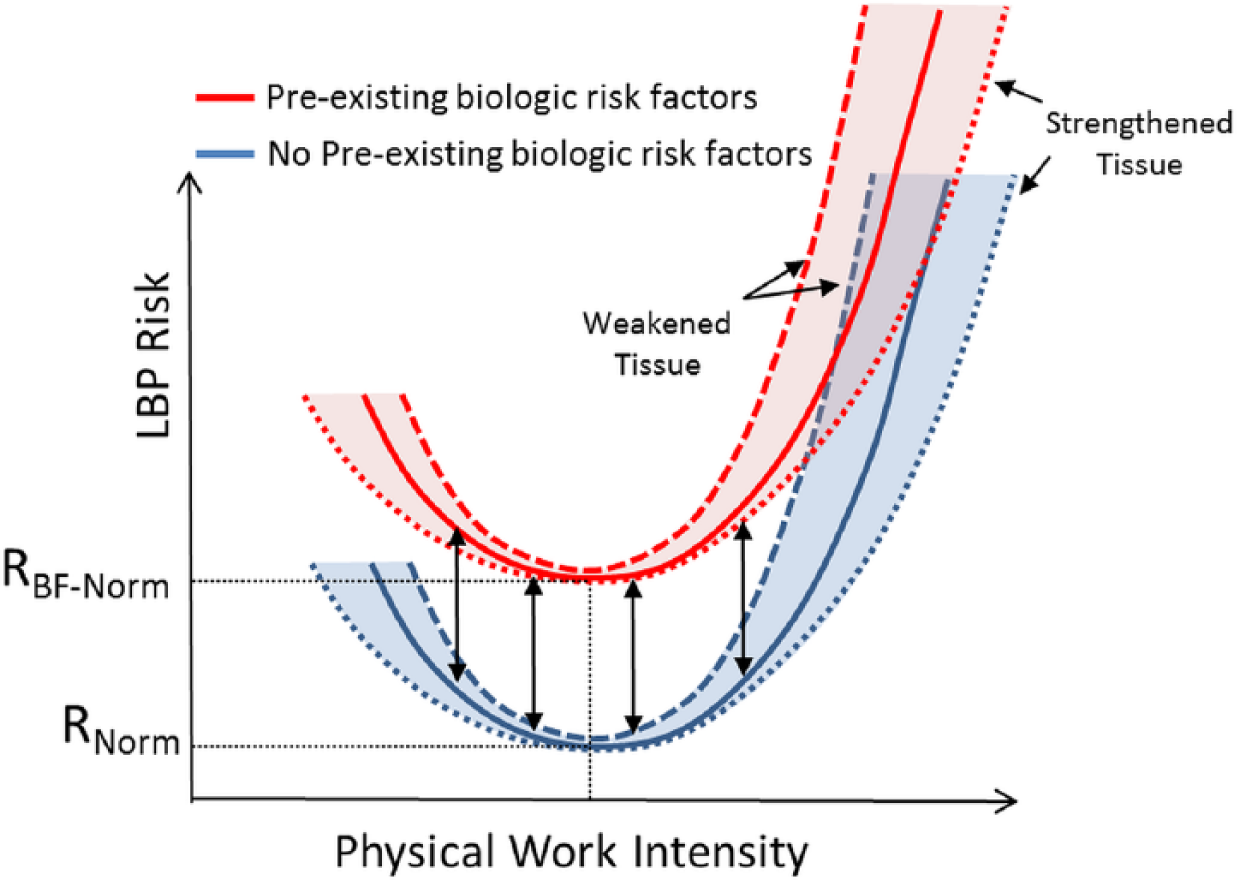
Preexisting biologic factors influence risk. A preexisting biologic risk factor, such as tissue damage, inflammation, or genetic mutation, could weaken or sensitize the tissue. Loading applied to this weakened or sensitized tissue would be at a greater risk (RBF-Norm) of experiencing low back pain (LBP) compared to an unaffected individual (RNorm) at the same physical work intensity.
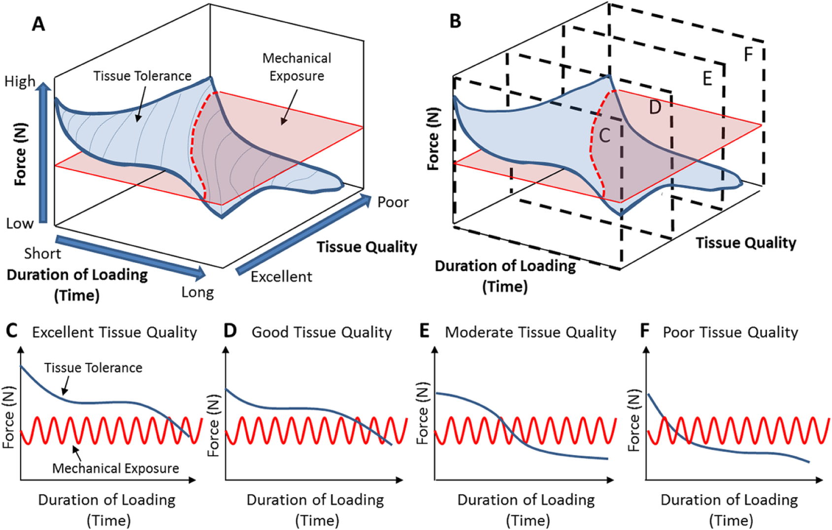
Tissue tolerance is a function of multiple factors. (A) Conceptual demonstration that the tissue tolerance is a function of duration of loading, the quality of the tissue, and its relationship with mechanical exposure. (B–F) Conceptual relationship between tissue tolerance and duration of loading at different tissue qualities. An individual is a at greater risk of tissue damage when the mechanical exposure is greater than the tissue tolerance. This relationship suggests that the risk of injury increases with time as tissue quality decreases.
Another way to incorporate mechano-biologic interactions into a preventive strategy to reduce workplace injuries may be to develop workplace routines that maintain loading within a “physiologic range” that promotes tissue strengthening or maintenance. This would require future research to identify the range of loading parameters (e.g., magnitude, frequency, and duration) that promotes matrix strengthening in the many different spinal tissues. In addition, this approach would require the use of personalized mathematical models that allow the calculation of the loads that the various tissues experience in different percentiles of the general population. Another potential application would be developing work routines or schedules that account for the time-dependent changes in spinal mechanics; for example, the incorporation of resting periods in between tasks that would allow time for recovery or alternating between high and low force exposures at different times of the day when the loads are borne by various spinal tissues.
Conclusion
In order to develop more universal tissue tolerances and understand their relationship to low back pain, it is important to recognize that both mechanical and biological factors influence the risk of experiencing low back pain. We propose that these mechano-biologic interactions are particularly relevant to both the perception of pain and the understanding that a tissue’s tolerance is dynamic and a function of multiple factors, including underlying pathology and time. Overall, the appreciation that the body is a system with many interacting factors may provide a more useful paradigm in the development of universal tolerance limits that approach the in vivo situation.
Key Points
Interactions between mechanical loading and biology can influence the perception of pain and regulates tissue strengthening and weakening.
A tissue’s tolerance is dynamic and is a function of multiple factors, including underlying pathology and time.
Both mechanical and biological factors influence the risk of experiencing low back pain, and as such, both factors should be incorporated into preventative strategies.
Footnotes
William S. Marras is the Honda Chair Professor in the Integrated Systems Engineering Department and director of the Spine Research Institute at The Ohio State University. He received his PhD in bioengineering from Wayne State University in 1982.
Benjamin A. Walter is a postdoctoral researcher in the Spine Research Institute and the Department of Biomedical Engineering at The Ohio State University. He received his PhD in biomedical engineering from The City College of New York in 2015.
Devina Purmessur is an assistant professor in the Department of Biomedical Engineering at The Ohio State University. She received her PhD in molecular pathology from the University of Manchester in 2008.
Prasath Mageswaran is a research engineer in the Spine Research Institute at The Ohio State University. He received his PhD in applied biomedical engineering from Cleveland State University in 2012.
Matthew G. Wiet is a master’s student in the Department of Biomedical Engineering at The Ohio State University. He received his bachelor’s degree in molecular, cell and developmental biology from the University of California Los Angeles in 2013.