
Abstract
Objective
Telangiectasias of the lower legs are intradermal dilatations of the subpapillary venous plexus, but their pathophysiology and risk factors are still largely unknown. The purpose of this review is to summarize the current knowledge on the pathophysiology and risk factors for telangiectasias.
Methods
A systematic review of the literature indexed in Medline completed with textbooks and European phlebology journals from the French, Swiss, and German phlebology societies was performed.
Results
A multitude of risk factors and several pathophysiological hypotheses, such as reflux, arterio-venous micro-shunts, parietal, and connective tissue abnormalities, are described in the literature. The different hypotheses are discussed and put in a clinical perspective, in particular their therapeutic implications for phlebologists.
Conclusion
In conclusion, pathophysiology and risk factors of telangiectasias are still largely unknown, and a better understanding could improve treatment results and reduce recurrence.
Introduction
Telangiectasias form a heterogeneous group including many types and affecting different parts of the body. They can be limited to the face like in couperose, or they can cover several parts of the body when they are associated with systemic diseases like collagenosis, cirrhosis, or congenital vascular malformations. On the lower legs, however, most telangiectasias are related to chronic venous disorders, and the present review will be limited to the latter group.
Telangiectasias of the lower leg measure less than 1 mm diameter and are defined as C1 in the CEAP classification. 1 Aesthetic concern is the most frequent reason for patient's consultation. Except for perimalleolar telangiectasias, known as corona phlebectatica paraplantaris, which is a sign of chronic venous insufficiency, telangiectasias of the lower leg (C1 A or S E P A S P 1N) represent a minor medical problem regarding prognosis and symptoms, but remain a challenge for the aesthetical treatment. Better knowledge of the pathophysiological mechanism and risk factors could improve treatment results.
Methodology
This review is based on an extensive Medline research completed with information from textbooks and European phlebology journals from the French, Swiss, and German phlebology societies, which are not indexed in Medline.
Epidemiology, symptoms, and quality of life
The prevalence of the telangiectasias in the general population is high and varies between 60 and 86% as they are isolated or associated with varicose veins (C2 according to CEAP classification).2,3 Their frequency increases with the age. 2 Medium age for isolated telangiectasias is 47 years, and it is 57.4 years when they are associated with varicose veins (C2). A linear relation exists between the number of telangiectasias and varicose veins; the more telangiectasias are present, the higher is the probability of having varicose veins. In the Edinburgh Vein Study, 2 telangiectasias were more frequent in women than in man, in a ratio of 4 to 3. But this was not the case in the Bonn Vein Study, 3 where their prevalence was identical in both sexes.
In the literature, the relation between telangiectasias and venous symptoms is contradictory.
Kröger et al. examined 9100 persons and observed that C1 patients presented more often feeling of swelling, muscular cramps, and restless leg symptoms 4 than the control group. In the Edinburgh Vein Study, 5 a significant difference of symptoms of heavy legs and edema was found only in women. In the German population, as many as 11.4% of the patients presenting isolated telangiectasias complained about edema. Only 0.2% presented cutaneous alterations like pigmentations. 3 In a selected population of patients treated with sclerotherapy, 53% described pain and a burning sensation in the territory of telangiectasias. After treatment, symptoms disappeared in 85% of the patients. 6 However, the San Diego study 7 did not find any difference in the frequency of symptoms between the class C1 and the control group.
The presence of telangiectasias does not significantly modify general quality of life (QoL; SF36 questionnaire). In the Bonn Vein Study, 3 a progressive reduction of the physical QoL was observed through the different CEAP classes, from 54.2 (class C0) to 40.7 (class C5). Telangiectasias obtained 51.5, as varicose veins C2 obtained a score of 48.2, which is a little bit lower than that of the general population (50). No difference in the psychological score was observed. In selected patients 8 consulting for an aesthetical treatment for their telangiectasias, physical score was normal 54.1. But the psychological score (48.5) was lower than that of the general population, probably because they were aesthetically affected.
Histology
Telangiectasias of the lower legs are intradermal dilatations of the subpapillary venous plexus. Their vein wall is often asymmetrically thickened, containing collagen, muscular, and more rarely elastic fibers. Under electron microscopy, altered and dysplastic collagen can be observed. 9 Chanvallon et al. 10 found two kinds of telangiectasias: The ones related to an insufficient vein were dilated, but conserved a normal vein wall structure, without any irregularities of the media. On the other hand, isolated telangiectasias (without associated insufficient veins) were tortuous, with variable thickness of the muscular media, suggestive of a hamartoma.
In atypical telangiectasias, a biopsy should be performed. Thickened wall of the dilated telangiectasias containing hyaline material positive for collagen IV at immunohistochemistry is typical for cutaneous collagenous vasculopathy (CCV). 11 It is a rare microangiopathy of unknown origin, which develops in middle-aged patients. Like generalized essential telangiectasias (GET), it usually appears on the lower leg before ascending gradually and symmetrically on the trunk, upper arms. 12 CCV may be successfully treated with a combination of optimized pulsed light and vascular laser. 11 GET, also from unknown etiology, is mainly a clinical diagnosis. Histology reveals fine-wall telangiectasias with flattened endothelial cells with usually no dermal or epidermal abnormalities. 12 There have been reports of treatment success with tetracycline, ketoconazole, acyclovir, sclerotherapy, and vascular laser therapy. 13
Pathophysiological hypothesis
Pathophysiology of telangiectasias of the lower legs is different from that of varicose veins and remains still a mystery. Several types of telangiectasias and pathophysiological mechanism exist and can be found on the same leg. Different theories have been proposed and are discussed in the following section.
The Reflux theory: Telangiectasias are secondary to venous reflux and venous hypertension
Arguments for
Several Doppler and venographic studies found an association between varicose veins and telangiectasias. In C1 patients, presenting only telangiectasias, Thibault et al. 14 found a saphenous reflux on Duplex ultrasound in 28% of the patients; and Engelhorn et al. even in 46% of the patients. 15 Analysis of the segmental distribution of the reflux in the great saphenous vein showed that the reflux was predominant in the calf segment (56%), it was rare at the sapheno-femoral junction (5%). 16 An ascending venography study was performed in 15 patients presenting telangiectasias at the lateral aspect of the thigh. 17 In eight cases, a direct relation between a major branch of the telangiectasias and the saphenous trunk was found, four of which had an incompetent saphenous vein. In two cases, a vein drained directly in the femoral vein through a perforator. In five cases, contrast diffused through the telangiectasias without any specific drainage. Performing the technique of micro-venography, Blin 18 discovered communications between telangiectasias and the deep venous system through small perforators. Unfortunately, his study was not able to determine the direction of blood flow or to specify if these micro-perforating veins were in- or out-flow pathways, neither their physiological nor pathological function. Albanese et al. 19 described already a “subdermic lateral venous system” of the thigh and calf. This system may become varicose even in the absence of incompetent saphenous veins. It presents multiple connections with the deep veins through small perforators located near the knee. These could serve as the primary source of reflux to transmit venous hypertension. Several authors14,20,21 described a relation between incompetent perforators and telangiectasias. Schuller-Petrovic et al. 22 found that resistant telangiectasias to sclerotherapy were commonly connected to a perforating vein. In 26 patients without deep and superficial venous insufficiency, refractory to at least three sessions of sclerotherapy, a feeding perforating vein was found in 14 patients. A direct connection with the great, small or anterior accessory saphenous veins was found in six patients. No connection was detected on Duplex ultrasound in the remaining six patients. The perforators and feeding veins were treated with ultrasound-guided foam sclerotherapy (polidocanol 0.5%). At three months, telangiectasias were either completely or at more than 75% eradicated, without any recurrence at 14 months' follow-up. This study demonstrates very well the relation between feeding veins and perforators and how important their eradication is to ensure a satisfactory treatment result of the telangiectasias.
Several studies showed a relation between insufficient reticular veins and telangiectasias.
Both Somjen et al. 21 and Weiss et al. 23 found reflux in 88% of the feeding reticular veins on Doppler examination. Raymond-Martinbeau et al. 24 found a direct relation between reticular veins and telangiectasias in 71% of the cases; and Mariani et al. 25 even found connections in 100% of the 200 areas of telangiectasias examined with side-illumination and Doppler. In a duplex ultrasonographic study of 43 asymptomatic limbs with lateral thigh telangiectasias, 26 all limbs had incompetent reticular veins. Twenty incompetent perforators were connected to the reticular veins. Perforator location was: lateral thigh 4, posterior thigh 2, lateral knee 4, lateral leg 8, posterior leg 2. In 10 limbs, reticular veins were not connected to perforators. In a post-mortem study, Wienert et al. 27 injected in 35 skin-muscle tissue blocks highly polymerizing plastic. A total of 28 areas of telangiectasias could be analyzed by electron microscopy, which revealed dilated venules and incompetent terminal valves in the subpapillary venous plexus.
Careful dissection by Taylor et al. 28 of superficial veins revealed a large number of valveless veins, which linked adjacent valved venous territories. They called them oscillating veins and supposed that these oscillating veins could allow equilibration of flow and pressure throughout the tissue and play a rule in thermoregulation. Green 20 postulates that these oscillating veins explain the confusion and disagreement about the apparent incompetence of the reticular veins.
Clinically, we can observe perimalleolar telangiectasias (corona phlebectatica paraplantaris) in case of deep venous insufficiency, post-thrombotic syndrome, saphenous vein and leg perforator incompetence. Therefore, a relation between reflux and telangiectasias does clearly exist. Widmer classified the corona phlebectatica as the first stage of chronic venous insufficiency. The corona phlebectatica was until now not included as a clinical sign in the CEAP classification. 1
Arguments against
In the Edinburgh Vein Study, most telangiectasias were localized at the thigh or the knee level. Only a minority was present on the lower third of the leg. These localizations are in contrast to the distal localization of chronic venous disease (CVD) skin modifications induced by venous hypertension. Pathophysiology of telangiectasias seems therefore to be different from CVD. In the typical clinical maritime pine presentation (Figure 1), telangiectasias of the lateral aspect of the thigh present an inverted pressure gradient to the feeding reticular veins. It is questionable if the venous hypertension can be provoked by reflux from distally located reticular veins or perforators and if it can be higher than the pressure gradient provoked by ground gravity, which is as high as 85 mmHg at the ankle in standing position?
29
Except in the case of a saphenous anterior accessory vein, or an incompetent perforating vein of the proximal thigh, reticular veins of the lateral aspect of the thigh drain mainly distally through perforating veins located near the knee or the leg.
26
Opposite to the static pressure model of gravity, we postulate a more dynamic venous system. During venous systole provoked by the very high pressure induced by muscular contraction, venous pressure spreads through incompetent micro-perforators throughout the venous system and more particularly to the weakest resistant veins, even if they go proximally. As a consequence, venous pressure rises in the venous columns, telangiectasias and reticular veins dilate, as they cannot properly drain against this pressure gradient. Only secondarily, the valves of the feeding reticular vein become incompetent, and reflux is detected on Duplex ultrasound. Therefore, dynamic systolic venous hypertension induced by an incompetent draining perforator and not reflux itself is likely to be at the origin of the pathological process. Garde
30
proposed a different hypothesis. He suggested that the reticular varicose veins and the telangiectasias of the lateral aspect of the thigh belong to the territory of the small saphenous vein and that they are secondary to the high compressive stress that experiences the sapheno-popliteal junction.
Maritime pine. The feeding reticular vein is located distally to the telangiectasias.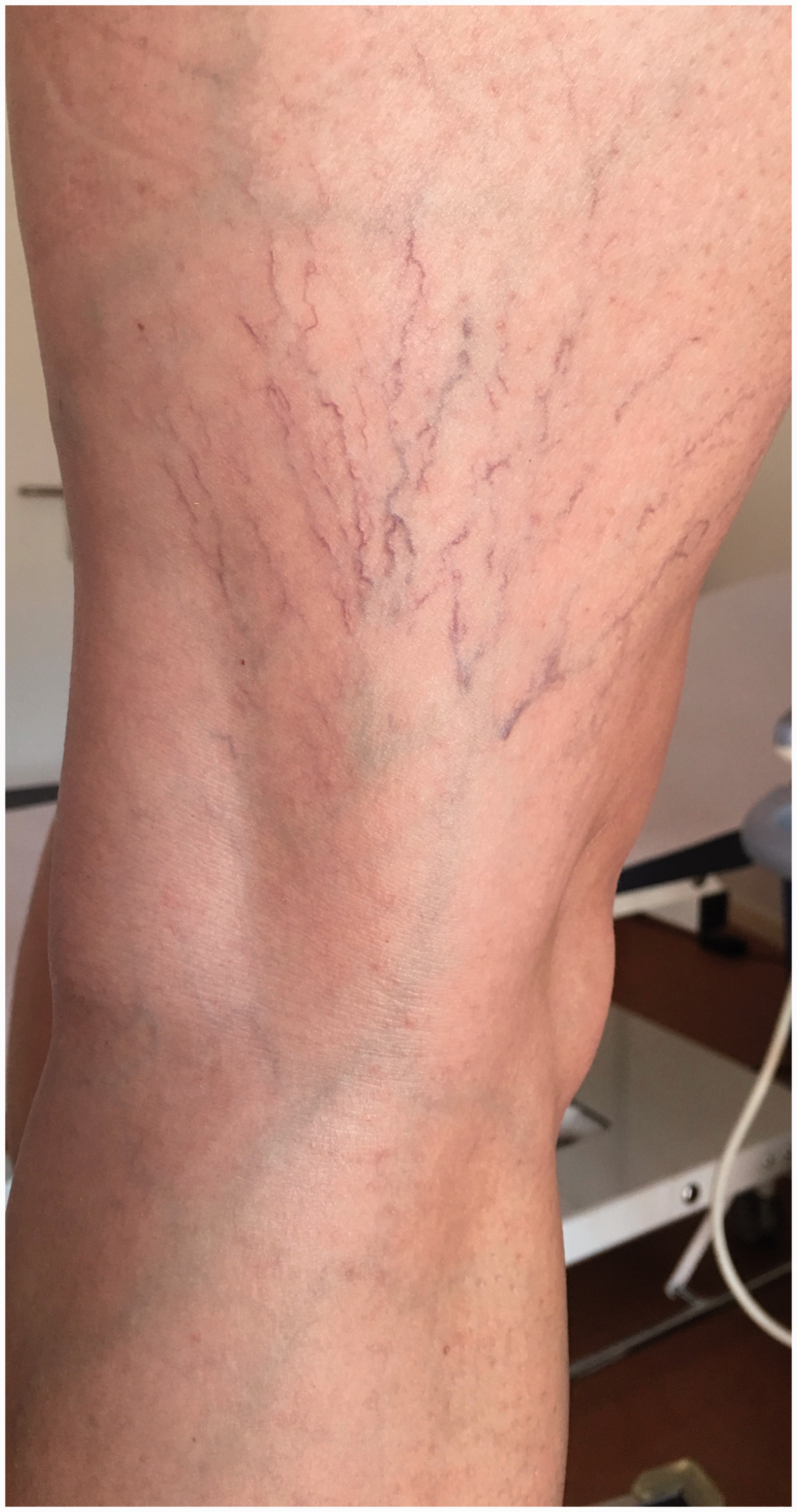
In conclusion, even if a clear relation exists between telangiectasias and reflux originating from perforators, saphenous and reticular veins, this pathophysiological mechanism cannot explain all telangiectasias. Particularly, the pathophysiological process of the reticular varicose veins and the telangiectasias of the lateral aspect of the thigh remain unclear. As reflux can hardly go upwards against gravity, we suggest that dilatation of reticular veins and telangiectasias are a consequence of dynamic systolic venous hypertension through incompetent distal perforating veins.
Any source of reflux must be suppressed to ensure treatment success. 22 Because of the proven relation of some telangiectasias and incompetent feeding veins, it is suggested to perform a Duplex ultrasound examination before treating telangiecasias. But in case of unsuccessful sclerotherapy, a Duplex ultrasound is strongly recommended prior to further treatment. Similarly, as we know from clinical experience of sclerotherapy and ambulatory hook phlebectomy, suppression of the incompetent perforators and the feeding reticular vein of the lateral aspect of the thigh will remove hypertension in the telangiectasias and resolve them. In case of phlebectomy, it is important to make the incision at each curve of the vein, as it is then often in relation with a perforator or a communicating vein, as described by Albanese et al. 19 and clinically demonstrated by Ramelet. 31
The micro-shunt theory
On 26 biopsies of telangiectasias, De Faria et al. 32 found one arterio-venous anastomosis. When a biopsy is performed in the presence of a pulsatile Doppler flow next to the telangiectasias, as many as 16 arterio-venous shunts/18 biopsies can be found. 33 In these cases, Bihari et al. 34 found not only the feeding arteriole but also an arterialization of the draining vein and venules. They also observed a significantly higher flow in telangiectasias in comparison to the surrounding skin (5 areas of telangiectasias/22 legs). They concluded that this higher flow has to be provoked by an arterio-venous shunt. Goldman et al. 35 excised five ulcerations, which occurred after sclerotherapy of telangiectasias. Neither the sclerosing drug (polidocanol) nor its concentration (0.5% liquid) could explain these ulcerations, as at this concentration polidocanol does not provoke any necrosis even with direct injection into the skin. In all five cases, they found an occluded underlying arteriole. They concluded that the sclerosing agent diffused through an arteriolo-venous micro-shunt. Regarding the technique used, an accidental intra-arteriolar injection seems implausible. Biegeleisen 36 formulated the hypothesis that matting (new fine red telangiectasias which arise shortly after sclerotherapy) is explained by unclosure of an arteriolo-venous micro-shunt. He suggested that the sclerosing agent is strong enough to close the telangiectasias but not for the arteriolo-venous micro-shunt. As consequence, the treated territory remains fed by a small arteriole, which promotes development of matting by redistribution of the blood flow.
In conclusion, the risk of ulceration is very small (<1% 35 ) if sclerotherapy is performed with mild agents (polidocanol 0.25- 0.5%, chromated glycerin), which do not provoke any necrosis even if injected accidentally into skin.35,37 If only areas with negative Doppler examination are treated (in Bihari's study, 33 69% of the areas were Doppler positive in C1 patients presenting only telangiectasias without any sign of CVD), we would have to avoid treating most of the territories. Anatomical, histological, venographic studies are limited by a too small number of cases to permit any conclusions. De Faria et al. 32 found only one arteriolovenous micro-shunt in 26 biopsies, Wienert et al 27 did not found any in 28 biopsies, as did not Böhler-Sommeregger et al. 17 who studied only 15 venography cases.
Even if arteriolo-venous micro-shunt can explain some ulceration after sclerotherapy, the risk is minimal. It is important to adopt a cautious injection technique with injection of only small volumes with a minimal injection pressure to avoid the risk of a possible passage of the sclerosing agent through an arteriolo-venous micro-shunt and the occlusion of an adjacent arteriole. Moreover with this injection technique, we limit inflammatory response, which can be responsible for matting and pigmentation. In current practice, arteriolo-venous micro-shunt can therefore be ignored. But they could explain some treatment failures (Figure 2). It is interesting to note that telangiectasias are present in the Klippel-Trenaunay syndrome, a combined vascular malformation associated with limb overgrowth (International Society for the Study of Vascular Malformations, ISSVA classification 2014). Pathophysiology of limb hypertrophy is still under debate. It could be secondary to active or passive hyperemia, but also to microscopic arterio-venous shunts.
38
In this situation, are the telangiectasias dysplastic veins, simply dilated veins secondary to the varicose veins or are they related to the arteriolovenous micro-shunt?
In this blow-out of red telangiectasias, when we apply a finger pressure in the middle of the spot, telangiectasias fills immediately again from the center with a centrifugal flow. In these situations, I am very careful, as treatment results can be disappointing. An underlying arterio-venous micro-shunt or a microperforator can be suspected.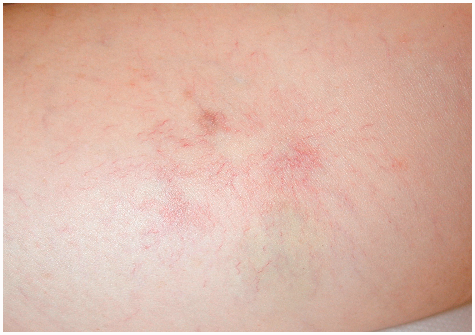
The inflammatory and parietal theory
The hypothesis of a primary default of the vein wall was proposed: Primary venule dilatation provokes blood stagnation, which could lead to leucocyte adhesion and vein wall alterations. The consequence is further dilatation and worsening of the telangiectasias. 39 But are there any consequences for the treatment?
The connective and adipose tissue theory
Fat lobules could reduce venous return, provoke localized venous hypertension, and telangiectasias. It is an attractive hypothesis, which needs further research. Would there be a link between lipodystrophy and telangiectasias? 40
The red and blue telangiectasias
Red telangiectasias are classically more resistant to sclerotherapy than the blue ones. Different hypothesis were published. Biegeleisen 36 suggested that the red telangiectasias were a consequence of small arteriolo-venous shunts, which provokes high inflow. This permanent flow would be responsible for the resistance to sclerotherapy. Sommer et al. 41 supposed that the red telangiectasias represent the arterial loop of the capillaries and the blue ones the venous loop. They thought that a slight pressure increase in the venous system would lead to a backflow of venous blood in the arterial loop of the capillary, with a consecutive dilatation of the arterial loop, which thereafter gets visible as a red telangiectasia. To support their theory, they measured the difference of oxygenation in the telangiectasias. Red telangiectasias had an average oxygen concentration of 5.9 kPa (44.25 mmHg), while it was only 5.11 kPa (38.3 mmHg) in the blue ones. But in my opinion, this very small difference speaks against an arterial contamination of venous blood, as the pO2 is usually as high as 11 kPa (82.5 mmHg) in the arteries and 4 kPa (30 mmHg) in the veins.
Wienert et al.
27
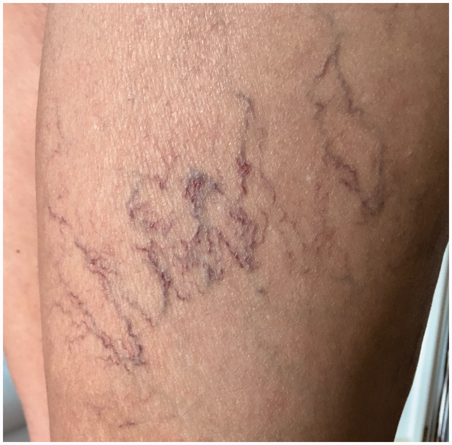
gave the answer to this question in their post-mortem electron microscopy study. They could clearly demonstrate that both were veins. Venous vessels have a rounded, polygonal pattern of endothelial cells orientated independently of the direction of the vessel. On the other hand, arterial vessels are characterized by elongated, streamlined endothelial cells running parallel to the vessel's direction. Both the red and the blue telangiectasias have the specific polygonal endothelial cell pattern of veins. Both also have multiple connections with the subpapillary venous plexus. The only difference they noticed was the diameter: Red telangiectasias (Figure 3) have a smaller diameter (0.227 mm) than the blue (0.435 mm) (Figure 4). Depth of telangiectasias in the skin was unfortunately not measured in this study, although depth could also partly explain color difference (red telangiectasias are more superficial).
Red telangiectasias. Blue telangiectasias. They have only a greater diameter than the red one. They can get as much dilated that they can become telangiectactic pearls, which have a risk of hemorrhagic rupture.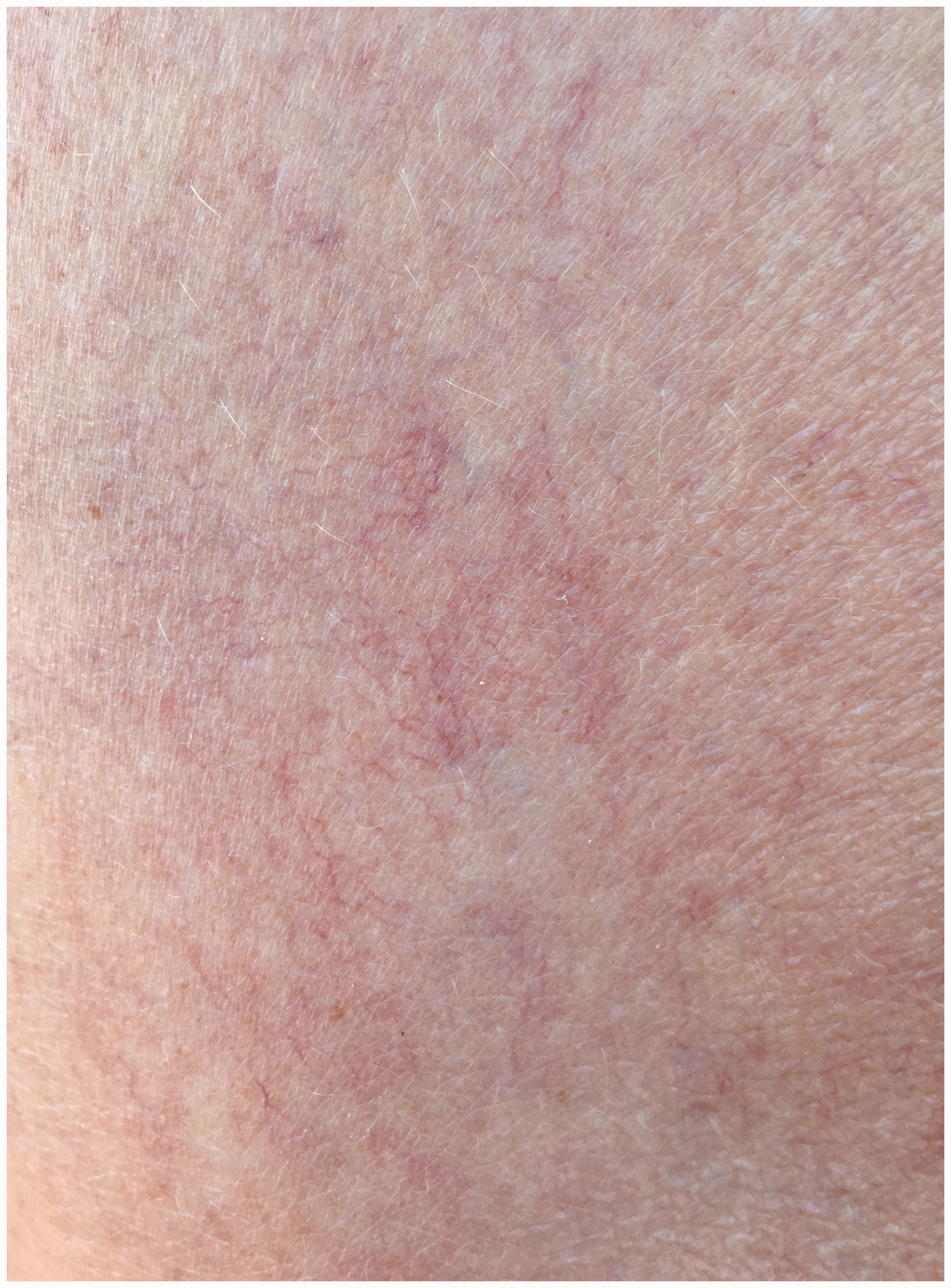
As already mentioned, GET can start clinically at the ankles with fine red telangiectasias (red sock-like appearance at the ankles). In atypical telangiectasias, this entity must be suspected, and a biopsy with immunohistochemistry must be performed to distinguish GET and CCV. 12
In conclusion, red telangiectasias are definitively veins, as are the blue ones. Why they are more difficult to treat and why they respond less to sclerotherapy remains still unclear. In my opinion, red telangiectasias are just younger veins in a process of ageing which is stimulated by angiogenic or inflammatory mediators as observed in matting. Sclerotherapy closes the telangiectasias but could be ineffective against growth factors and angiogenesis, which still go on.
In my experience, red telangiectasias respond better to mild sclerosing agents like chromated glycerin or liquid polidocanol 0.25%. Using stronger sclerosing agents, large volumes or high injection pressure (for this reason I do not use smaller syringes than 2.5 ml, with smaller syringes producing higher injection pressure) enhances inflammatory response and risk of matting. Cutaneous laser could be an alternative for resistant red telangiectasias to sclerotherapy, as far as the feeding reticular varicose veins were previously treated.
Sclerotherapy remains the first choice technique, as the sclerosing agent has the advantage not only to treat the telangiectasias but also to be able to eliminate the reticular feeding vein, diffuse, and close communicating veins or micro-perforators.
Risk factors
Age
Age is certainly the most important risk factor. Prevalence of telangiectasias increases progressively with age. 2
Heredity
The Edinburgh Vein Study 2 could also confirm the importance of a positive familial history of telangiectasias as a risk factor. Fifty-four percent of the C1 patients had a positive familial history of telangiectasias, compared to only 32% in the control group.
Hormones – Pregnancy
Estrogen and progesterone hormones cause dilatation of the vein wall. 42 Progesterone provokes vasoplegia and increases permeability of capillaries. It multiplies the number of and dilates venules. Estrogen denatures collagen of the media and stimulates angiogenic growth factors. One study found estrogen and progesterone receptors in endothelial cells. 43 But Sadick et al. did not find any in telangiectasias, 44 or in matting areas. 45 An argument for hormonal influence is that telangiectasias can develop during pregnancy and then regress after birth. This argument is supported in the case of telangiectasias appearing at the beginning of pregnancy, where venous pelvic hypertension cannot yet be incriminated. In the epidemiological study by Ruckley et al., 2 only 44% of the women presenting isolated C1 telangiectasias have taken the contraceptive pill, but surprisingly, 84.4% of the women in the control group have taken the contraceptive pill. These data suggest that contraceptive pill could have a protective effect. The same observation was true for hormone replacement treatment; 26.9% of the C1 patients never took any hormones, in comparison to only 2.2% in the control group. These surprising data could be biased because of the small number of patients in the C1 group (n = 29, versus 122 in the control group). On the other hand, these results could also be explained by the fact that women who had one or more pregnancies are likely to take less contraceptive pill than nulliparous. In contrast to the contraceptive pill, pregnancy was a very strong risk factor as 100% of the C1 women had at least one pregnancy, compared to only 40.5% of the women of the control group. This means that in the control group, the protective factor was mainly the absence of pregnancy instead of the contraceptive pill intake.
Sun exposure
A linear correlation exists between rate of sun exposure and telangiectasias. Sunlight degrades collagen and elastic fibers, located around telangiectasias. 42 In a national survey study to document deleterious effect of ultraviolet radiation on skin in the USA between 1971 and 1974, a significant correlation was observed between actinic skin damage (senile elastosis, actinic keratosis) and the presence of telangiectasias. Prevalence of telangiectasias was doubled in persons having high sunlight exposure and actinic skin lesions in comparison to those having low sun exposure. 46
Trauma
Patients often report that a localized trauma (e.g. a tennis ball hit) is at the origin of their telangiectactic spot. A localized trauma could initiate angiogenesis and telangiectasias. 42 Rupture of the vessel wall not only provokes a bruise but liberates also endothelial growth factors. Tissue hypoxia induced by the local compression from the hematoma, an elevated venous pressure in a reflux territory, a bad drainage and venous stasis are additional factors that stimulate angiogenesis. 30 The risk of developing telangiectasias after venous surgery (stripping, hook phlebectomy…) is well known. In this case, the same pathophysiologic process could provoke matting.
A localized trauma could also damage perforators, or create arteriolo-venous micro-shunts as source of secondary telangiectasias.
Standing position – Lifestyle
Standing position is a well-known risk factor for varicose veins C2.47–49 Unfortunately, in the published literature, the group of patient presenting only isolated telangiectasias C1 was not analyzed.
Obesity
In a sonographic study of thigh telangiectasias, 26 obese and overweight patients (BMI > 25) presented a significant higher prevalence of incompetent perforators and reticular veins, and had a larger diameter when compared to normal weight.
Iatrogenic risk factors
Prolonged use of oral steroids or application of strong topical steroids can induce telangiectasias, which are also observed in radiodermatitis. 35
Conclusion
The pathophysiology of telangiectasias remains mysterious. Several types of telangiectasias and several pathophysiological mechanisms exist. The majority of the telangiectasias are certainly secondary to reflux originating either from feeding reticular veins, from perforators, or varicose veins. The pathophysiological mechanism of telangiectasias of the lateral aspect of the thigh may be different. Due their distal drainage, it is unlikely that reflux occurs against ground gravity. It is, however, possible that the pressure in the lateral vein system is pathologically increased due to an insufficient drainage perforator, which through dynamic venous systole hypertension transmission leads to the dilatation of the reticular veins and telangiectasias.
Before treating telangiectasias, a clinical and duplex ultrasound examination permits to precise diagnosis, to classify the venous disease, and especially to detect any underlying venous reflux. To get the greatest chance of an effective treatment, every reflux, particularly all feeding reticular veins must be eliminated, either in advance or during the same treatment session.
Telangiectasias can be related to arteriolo-venous micro-shunts, but their real prevalence is unknown. Expect for the risk of focal treatment failure, their presence is in my opinion not relevant, as the absolute risk of post-sclerotherapeutic skin ulceration is very low when mild sclerosing agents and an appropriate injection technique is applied.
The main risk factors of telangiectasias are age, heredity, and pregnancy. The real risk of estrogen and progesterone hormones is not established.
Footnotes
Declaration of Conflicting Interests
The author(s) declared no potential conflicts of interest with respect to the research, authorship, and/or publication of this article.
Funding
The author(s) received no financial support for the research, authorship, and/or publication of this article.
Informed consent
Not applicable.
Ethical approval
Not applicable.
Guarantor
PK.
Contributorship
PK researched literature and wrote the manuscript.
Acknowledgements
I would like to thank Albert-Adrien Ramelet MD (Montcherand) and Rolf P. Engelberger MD (Fribourg) for the critical reading of the manuscript.